
Phospho-Nrf2 (Ser40) Rabbit Polyclonal Antibody, Unconjugated
Artikelnummer:
BYT-ORB6544
- Bilder (8)


| Artikelname: | Phospho-Nrf2 (Ser40) Rabbit Polyclonal Antibody, Unconjugated |
| Artikelnummer: | BYT-ORB6544 |
| Hersteller Artikelnummer: | orb6544 |
| Alternativnummer: | BYT-ORB6544-50,BYT-ORB6544-100,BYT-ORB6544-200 |
| Hersteller: | Biorbyt |
| Wirt: | Rabbit |
| Kategorie: | Antikörper |
| Applikation: | ICC, WB |
| Spezies Reaktivität: | Human |
| Immunogen: | KLH conjugated Synthesised phosphopeptide derived from human Nrf2 around the phosphorylation site of Ser40 DF(p-S)QR |
| Konjugation: | Unconjugated |
| Alternative Synonym: | NFE2L2 | NRF2 (p-S40), p-NRF2, phospho-NRF2, HEBP1, IMDDHH, NRF2, Nrf-2, NF2L2_HUMAN, NFE2L2, NF-E2-related factor 2, NFE2-related factor 2, Nuclear factor, erythroid derived 2, like 2, |
| Phospho-Nrf2 (Ser40) Rabbit Polyclonal Antibody |
| Application Verdünnung: | WB=1:500-2000, ICC/IF=1:100-500 |
| Anwendungsbeschreibung: | Modification: Phosphorylated |
|
|
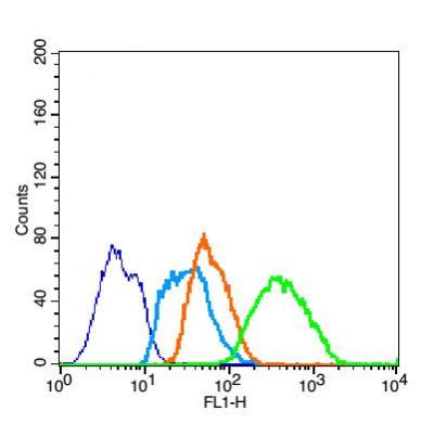
Blank control (blue line): Jurkat (fixed with 2% paraformaldehyde (10 min), then permeabilized with 90% ice-cold methanol for 30 min on ice). Primary Antibody (green line): Rabbit Anti-phospho-Nrf2 (Ser40) antibody (orb6544), dilution: 1 µg/10 6 cells, Isotype Control Antibody (orange line): Rabbit IgG. Secondary Antibody (white blue line): Goat anti-rabbit IgG-FITC, dilution: 1 µg/Test. |
|
|
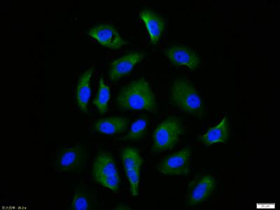
HepG2 cell, 4% Paraformaldehyde-fixed, Triton X-100 at room temperature for 20 min, Blocking buffer (normal goat serum) at 37C for 20 min, Antibody incubation with (phospho-Nrf2 (Ser40)) polyclonal Antibody, Unconjugated (orb6544) 1:100, 90 minutes at 37C, followed by a conjugated Goat Anti-Rabbit IgG antibody at 37C for 90 minutes, DAPI (blue) was used to stain the cell nuclei. |
|
|
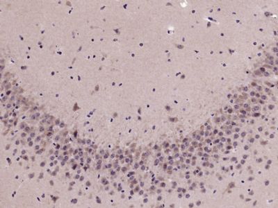
Paraformaldehyde-fixed, paraffin embedded (Mouse brain), Antigen retrieval by microwave in sodium citrate buffer (pH6.0), Block endogenous peroxidase by 3% hydrogen peroxide for 30 minutes, Blocking buffer (3% BSA) at RT for 30 min, Antibody incubation with (phospho-Nrf2 (Ser40)) Polyclonal Antibody, Unconjugated (orb6544) at 1:400 overnight at 4C, followed by conjugation to the secondary antibody (labeled with HRP) and DAB staining. |
|
|
Paraformaldehyde-fixed, paraffin embedded (Rat brain), Antigen retrieval by microwave in sodium citrate buffer (pH6.0), Block endogenous peroxidase by 3% hydrogen peroxide for 30 minutes, Blocking buffer (3% BSA) at RT for 30 min, Antibody incubation with (phospho-Nrf2 (Ser40)) Polyclonal Antibody, Unconjugated (orb6544) at 1:400 overnight at 4C, followed by conjugation to the secondary antibody (labeled with HRP) and DAB staining. |
|
|
Sample: Lane 1: Hela (Human) Cell Lysate at 30 ug, Lane 2: HepG2 (Human) Cell Lysate at 30 ug, Lane 3: MCF-7 (Human) Cell Lysate at 30 ug, Lane 4: Jurkat (Human) Cell Lysate at 30 ug, Lane 5: B16 (Human) Cell Lysate at 30 ug, Lane 6: A549 (Human) Cell Lysate at 30 ug, Primary: Anti-phospho-Nrf2 (Ser40) (orb6544) at 1/1000 dilution, Secondary: IRDye800CW Goat Anti-Rabbit IgG at 1/20000 dilution, Predicted band size: 68 kD, Observed band size: 135 kD. |
|
|
IHC-P image of Mouse brain tissue using Nrf2 (phospho-Ser40) antibody |
|
|
IF analysis of HepG2 cell lysate using Nrf2 (phospho-Ser40) antibody |
|
|
Flow cytometric analysis of Jurkat cell lysate using Nrf2 (phospho-Ser40) antibody |
Produktgarantie und fachkundiger Support
