
Phospho-P53 (Ser46) Rabbit Polyclonal Antibody, Unconjugated
Artikelnummer:
BYT-ORB6598
- Bilder (7)


| Artikelname: | Phospho-P53 (Ser46) Rabbit Polyclonal Antibody, Unconjugated |
| Artikelnummer: | BYT-ORB6598 |
| Hersteller Artikelnummer: | orb6598 |
| Alternativnummer: | BYT-ORB6598-50,BYT-ORB6598-100,BYT-ORB6598-200 |
| Hersteller: | Biorbyt |
| Wirt: | Rabbit |
| Kategorie: | Antikörper |
| Applikation: | FC, ICC, IF, IHC-Fr, IHC-P, WB |
| Spezies Reaktivität: | Human, Mouse |
| Immunogen: | KLH conjugated Synthesised phosphopeptide derived from human P53 around the phosphorylation site of Ser46 ML(p-S)PD |
| Konjugation: | Unconjugated |
| Alternative Synonym: | TP53 | p53 (p-S46), p-p53, phospho-p53, BCC7, BMFS5, LFS1, P53, TRP53, Tp53, bbl, bfy, bhy, p44, P53_BOVIN, Tumor suppressor p53, P53_HUMAN, Antigen NY-CO-13, Phosphoprotein p53, P53_MOUSE, P53_RAT, |
| Phospho-P53 (Ser46) Rabbit Polyclonal Antibody |
| Klonalität: | Polyclonal |
| Konzentration: | 1mg/ml |
| Molekulargewicht: | 43 kDa |
| UniProt: | P04637 |
| Puffer: | 0.01M TBS (pH7.4) with 1% rAlbumin, 0.02% Proclin300 and 50% Glycerol. |
| Formulierung: | Liquid |
| Target-Kategorie: | TP53 |
| Application Verdünnung: | WB=1:500-2000, IHC-P=1:100-500, IHC-F=1:100-500, ICC/IF=1:100-500, IF=1:100-500, Flow-Cyt=0.2µg /test |
| Anwendungsbeschreibung: | Modification: Phosphorylated |
|
|
A549 cell, 4% Paraformaldehyde-fixed, Triton X-100 at room temperature for 20 min, Blocking buffer (normal goat serum) at 37C for 20 min, Antibody incubation with (phospho-P53 (Ser46)) polyclonal Antibody, Unconjugated (orb6598) 1:100, 90 minutes at 37C, followed by a conjugated Goat Anti-Rabbit IgG antibody at 37C for 90 minutes, DAPI (blue) was used to stain the cell nuclei. |
|
|
Blank control (blue line): Hela (fixed with 70% ethanol (Overnight at 4C) and then permeabilized with 90% ice-cold methanol for 30 min on ice). Primary Antibody (green line): Rabbit Anti-phospho-P53 (Ser46) antibody (orb6598), dilution: 0.2 µg/10 6 cells, Isotype Control Antibody (orange line): Rabbit IgG. Secondary Antibody (white blue line): Goat anti-rabbit IgG-FITC, dilution: 1 µg/Test. |
|
|
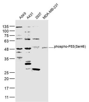
Sample: A549 (Human) Cell Lysate at 30 ug, A431 (Human) Cell Lysate at 30 ug, 293T (Human) Cell Lysate at 30 ug, MDA-MB-231 (Human) Cell Lysate at 30 ug, Primary: Anti-phospho-P53 (Ser46) (orb6598) at 1/300 dilution, Secondary: IRDye800CW Goat Anti-Rabbit IgG at 1/20000 dilution, Predicted band size: 43 kD, Observed band size: 48 kD. |
|
|
Sample: Lane 1: A431 (Human) Cell Lysate at 30 ug, Lane 2: A549 (Human) Cell Lysate at 30 ug, Lane 3: MCF-7 (Human) Cell Lysate at 30 ug, Primary: Anti-phospho-P53 (Ser46) (orb6598) at 1/1000 dilution, Secondary: IRDye800CW Goat Anti-Rabbit IgG at 1/20000 dilution, Predicted band size: 53 kD, Observed band size: 55 kD. |
|
|
Sample: MCF-7 (Human) Cell Lysate at 30 ug, Primary: Anti-phospho-P53 (Ser46) (orb6598) at 1/300 dilution, Secondary: IRDye800CW Goat Anti-Rabbit IgG at 1/20000 dilution, Predicted band size: 43 kD, Observed band size: 48 kD. |
|
|
Tissue/Cell: human laryngocarcinoma, 4% Paraformaldehyde-fixed and paraffin-embedded, Antigen retrieval: citrate buffer (0.01M, pH6.0), Boiling bathing for 15 min, Block endogenous peroxidase by 3% Hydrogen peroxide for 30 min, Blocking buffer (normal goat serum) at 37C for 20 min, Incubation: Anti-phospho-P53 (Ser46) Polyclonal Antibody, Unconjugated (orb6598) 1:200, overnight at 4C, followed by conjugation to the secondary antibody and DAB staining. |
Produktgarantie und fachkundiger Support
