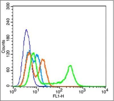
Flow cytometric analysis of EC9706 Cell using AKT1 (phospho-Thr34) antibody.
Western blot analysis of mouse embryos lysate using AKT1 (phospho-Thr34) antibody.
Blank control (blue): EC9706 (fixed with 2% paraformaldehyde for 10 min at 37C). Primary Antibody: Rabbit Anti-phospho-AKT1 (Thr34) antibody (orb6783, Green), dilution: 3 µg in 100 µl 1X PBS containing 0.5% BSA, Isotype Control Antibody: Rabbit IgG (orange), used under the same conditions, Secondary Antibody: Goat anti-rabbit IgG-FITC (white blue), dilution: 1:200 in 1X PBS containing 0.5% BSA.
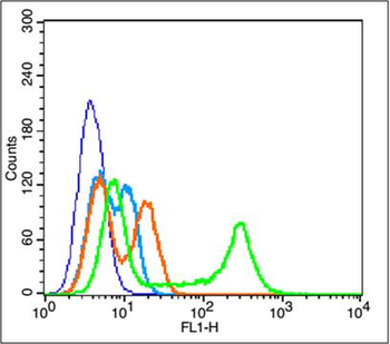
Blank control: A549. Primary Antibody (green line): Rabbit Anti-phospho-AKT1 (Thr34) antibody (orb6783), dilution: 2 µg/10 6 cells, Isotype Control Antibody (orange line): Rabbit IgG. Secondary Antibody: Goat anti-rabbit IgG-FITC, dilution: 1 µg/Test. Protocol, The cells were incubated in 5% BSA to block non-specific protein-protein interactions for 30 min at room temperature. Cells stained with Primary Antibody for 30 min at room temperature. The secondary antibody used for 40 min at room temperature. Acquisition of 20000 events was performed.
Paraformaldehyde-fixed, paraffin embedded (Mouse brain), Antigen retrieval by boiling in sodium citrate buffer (pH6.0) for 15 min, Block endogenous peroxidase by 3% hydrogen peroxide for 20 minutes, Blocking buffer (normal goat serum) at 37C for 30 min, Antibody incubation with (phospho-AKT1 (Thr34)) Polyclonal Antibody, Unconjugated (orb6783) at 1:400 overnight at 4C, followed by operating according to SP Kit (Rabbit) instructionsand DAB staining.
Sample: Lane 1: SiHa (Human) Cell Lysate at 30 ug, Lane 2: NIH/3T3 (Mouse) Cell Lysate at 30 ug, Lane 3: Adrenal glands (Mouse) Lysate at 40 ug, Lane 4: Skeletal muscle (Mouse) Lysate at 40 ug, Lane 5: Ovary (Mouse) Lysate at 40 ug, Lane 6: Lung (Mouse) Lysate at 40 ug, Lane 7: Cerebrum (Mouse) Lysate at 40 ug.
* Mehrwertsteuer und Versandkosten nicht enthalten. Irrtümer und Preisänderungen vorbehalten