Each vial contains 4mg Trehalose, 0.9mg NaCl, 0.2mg Na2HPO4.
Formulierung:
Lyophilized
Target-Kategorie:
Scaffold protein ILK
Application Verdünnung:
Western blot, 0.1-0.25µg/ml, Human, Mouse, Rat Immunohistochemistry(Paraffin-embedded Section), 1-2 µg/ml, Human Immunocytochemistry/Immunofluorescence, 5µg/ml, Human ELISA, 0.1-0.5µg/ml, -
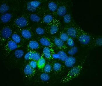
IF analysis of Integrin linked ILK using anti-Integrin linked ILK antibody. Integrin linked ILK was detected in immunocytochemical section of A431 cells. Enzyme antigen retrieval was performed using IHC enzyme antigen retrieval reagent for 15 mins. The cells were blocked with 10% goat serum. And then incubated with 5 µg/mL rabbit anti-Integrin linked ILK Antibody overnight at 4C. DyLight488 Conjugated Goat Anti-Rabbit IgG was used as secondary antibody at 1:100 dilution and incubated for 30 minutes at 37C. The section was counterstained with DAPI. Visualize using a fluorescence microscope and filter sets appropriate for the label used.
IHC analysis of Integrin linked ILK using anti-Integrin linked ILK antibody. Integrin linked ILK was detected in a paraffin-embedded section of human bladder epithelial carcinoma tissue. Heat mediated antigen retrieval was performed in EDTA buffer (pH8.0, epitope retrieval solution). The tissue section was blocked with 10% goat serum. The tissue section was then incubated with 2 µg/ml rabbit anti-Integrin linked ILK Antibody overnight at 4C. Biotinylated goat anti-rabbit IgG was used as secondary antibody and incubated for 30 minutes at 37C. The tissue section was developed using Strepavidin-Biotin-Complex (SABC) with DAB as the chromogen.
IHC analysis of Integrin linked ILK using anti-Integrin linked ILK antibody. Integrin linked ILK was detected in a paraffin-embedded section of human breast cancer tissue. Heat mediated antigen retrieval was performed in EDTA buffer (pH8.0, epitope retrieval solution). The tissue section was blocked with 10% goat serum. The tissue section was then incubated with 2 µg/ml rabbit anti-Integrin linked ILK Antibody overnight at 4C. Biotinylated goat anti-rabbit IgG was used as secondary antibody and incubated for 30 minutes at 37C. The tissue section was developed using Strepavidin-Biotin-Complex (SABC) with DAB as the chromogen.
IHC analysis of Integrin linked ILK using anti-Integrin linked ILK antibody. Integrin linked ILK was detected in a paraffin-embedded section of human laryngeal squamous cell carcinoma tissue. Heat mediated antigen retrieval was performed in EDTA buffer (pH8.0, epitope retrieval solution). The tissue section was blocked with 10% goat serum. The tissue section was then incubated with 2 µg/ml rabbit anti-Integrin linked ILK Antibody overnight at 4C. Biotinylated goat anti-rabbit IgG was used as secondary antibody and incubated for 30 minutes at 37C. The tissue section was developed using Strepavidin-Biotin-Complex (SABC) with DAB as the chromogen.
Western blot analysis of Integrin linked ILK using anti-Integrin linked ILK antibody. Electrophoresis was performed on a 5-20% SDS-PAGE gel at 70V (Stacking gel) / 90V (Resolving gel) for 2-3 hours. The sample well of each lane was loaded with 30 ug of sample under reducing conditions. Lane 1: human MCF-7 whole cell lysates, Lane 2: human Hela whole cell lysates, Lane 3: human U-87MG whhole cell lysates, Lane 4: human A431 whole cell lysates, Lane 5: human U20S whhole cell lysates, Lane 6: human PC-3 whhole cell lysates, Lane 7: rat heart tissue lysates, Lane 8: rat kidney tissue lysates, Lane 9: rat skeletal muscle tissue lysates, Lane 10: rat PC-12 whhole cell lysates, Lane 11: mouse heart tissue lysates, Lane 12: mouse kidney tissue lysates, Lane 13: mouse skeletal muscle tissue lysates, Lane 14: mouse C2C12 whhole cell lysates. After Electrophoresis, proteins were transferred to a Nitrocellulose membrane at 150 mA for 50-90 minutes. Blocked the membrane with 5% Non-fat Milk/ TBS for 1.5 hour at RT. The membrane was incubated with rabbit anti-Integrin linked ILK antigen affinity purified polyclonal antibody at 0.25 µg/mL overnight at 4C, then washed with TBS-0.1% Tween 3 times with 5 minutes each and probed with a goat anti-rabbit IgG-HRP secondary antibody at a dilution of 1:5000 for 1.5 hour at RT. The signal is developed using an Enhanced Chemiluminescent detection (ECL) kit with Ta
* Mehrwertsteuer und Versandkosten nicht enthalten. Irrtümer und Preisänderungen vorbehalten