Activated human peripheral blood mononuclear cells were used as the immunogen for the HLA-DRB1 antibody.
Konjugation:
Unconjugated
This mAb reacts with the beta-chain of HLA-DRB1 antigen, a member of MHC class II molecules. It does not cross react with HLA-DP and HLA-DQ. Its epitope is different from that of mAb L243. HLA-DR is a heterodimeric cell surface glycoprotein comprised of a 36kDa alpha (heavy) chain and a 28kDa beta (light) chain. It is expressed on B-cells, activated T-cells, monocytes/macrophages, dendritic cells and other non-professional APCs. In conjunction with the CD3/TCR complex and CD4 molecules, HLA-DR is critical for efficient peptide presentation to CD4+ T cells. It is an excellent histiocytic marker in paraffin sections producing intense cytoplasmic staining. True histiocytic neoplasms are similarly positive. HLA-DR antigens also occur on a variety of epithelial cells and their corresponding neoplastic counterparts. Loss of HLA-DR expression is related to tumor microenvironment and predicts adverse outcome in diffuse large B-cell lymphoma.
0.2 mg/ml in 1X PBS with 0.1 mg/ml rAlbumin and 0.05% sodium azide
Application Verdünnung:
Flow cytometry: 1-2ug/10 6 cells,Immunofluorescence: 2-4ug/ml,Western blot: 1-2ug/ml,Immunohistochemistry (FFPE): 0.25-0.5ug/ml for 30 min at RT
Anwendungsbeschreibung:
Application Notes: Optimal dilution of the HLA-DRB1 antibody should be determined by the researcher.1. Staining of formalin-fixed tissues is enhanced by boiling tissue sections in pH 9 10mM Tris with 1mM EDTA for 10-20 min followed by cooling at RT for 20 min2. The prediluted format is supplied in a dropper bottle and is optimized for use in IHC. After epitope retrieval step (if required), drip mAb solution onto the tissue section and incubate at RT for 30 min
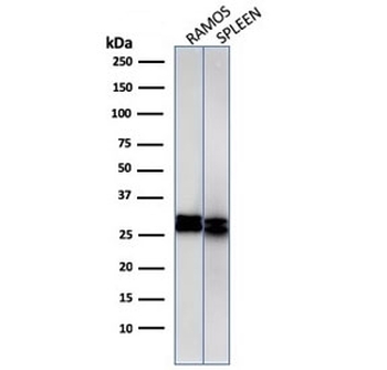
Western blot Anaysis of human 1) Ramos and 2) spleen cell Lyste using HLA-DRB1 antibody (clone HLA-DRB/1067). Predicted molecular weight ~30 kDa.
Western blot Anaysis of Ramos cell Lyste using HLA-DRB1 antibody (HLA-DRB/1067). Predicted molecular weight ~30 kDa.
IHC: Formalin-fixed, paraffin-embedded human Histiocytoma stained with HLA-DRB antibody (clone HLA-DRB/1067).
IHC: Formalin-fixed, paraffin-embedded human tonsil stained with HLA-DRB antibody (clone HLA-DRB/1067).
Immunofluorescent staining of Raji cells with HLA-DRB1 antibody (green, clone HLA-DRB/1067) and Reddot nuclear stain (red).
FACS staining of Raji cells with HLA-DRB1 antibody (clone HLA-DRB/1067), Red = isotype control, Blue = HLA-DRB1 antibody.
* Mehrwertsteuer und Versandkosten nicht enthalten. Irrtümer und Preisänderungen vorbehalten