A synthetic peptide corresponding to a sequence in the middle region of human YB1, identical to the related rat and mouse sequences.
Konjugation:
Unconjugated
Alternative Synonym:
Nuclease-sensitive element-binding protein 1, CCAAT-binding transcription factor I subunit A, CBF-A, DNA-binding protein B, DBPB, Enhancer factor I subunit A, EFI-A, Y-box transcription factor, Y-box-binding protein 1, YB-1, YBX1, NSEP1, YB1
Anti-YB1/YBX1 Antibody. Tested in Flow Cytometry, IHC, WB applications. This antibody reacts with Human, Mouse, Rat.
Klonalität:
Polyclonal
Konzentration:
Adding 0.2 ml of distilled water will yield a concentration of 500 µg/ml.
Each vial contains 4 mg Trehalose, 0.9 mg NaCl and 0.2 mg Na2HPO4.
Formulierung:
Lyophilized
Target-Kategorie:
Y-box-binding protein 1
Application Verdünnung:
Western blot, 0.1-0.5µg/ml, Human, Mouse, Rat Immunohistochemistry(Paraffin-embedded Section), 2-5 µg/ml, Human, Mouse, Rat Flow Cytometry (Fixed), 1-3 µg/1x10 6 cells, Human
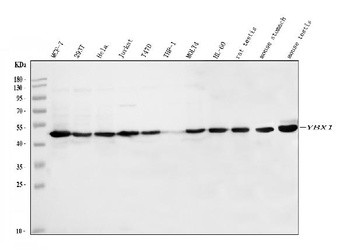
Western blot analysis of YBX1 using anti-YBX1 antibody (orb76271). Electrophoresis was performed on a 5-20% SDS-PAGE gel at 70V (Stacking gel)/90V (Resolving gel) for 2-3 hours. The sample well of each lane was loaded with 30 ug of sample under reducing conditions. Lane 1: human MCF-7 whole cell lysates, Lane 2: human 293T whole cell lysates, Lane 3: human HeLa whole cell lysates, Lane 4: human Jurkat whole cell lysates, Lane 5: human T47D whole cell lysates, Lane 6: human THP-1 whole cell lysates, Lane 7: human MOLT4 whole cell lysates, Lane 8: human HL-60 whole cell lysates, Lane 9: rat testis tissue lysates, Lane 10: mouse stomach tissue lysates, Lane 11: mouse testis tissue lysates. After electrophoresis, proteins were transferred to a nitrocellulose membrane at 150 mA for 50-90 minutes. Blocked the membrane with 5% non-fat milk/TBS for 1.5 hour at RT. The membrane was incubated with rabbit anti-YBX1 antigen affinity purified polyclonal antibody (Catalog orb76271) at 0.5 µg/mL overnight at 4C, then washed with TBS-0.1%Tween 3 times with 5 minutes each and probed with a goat anti-rabbit IgG-HRP secondary antibody at a dilution of 1:5000 for 1.5 hour at RT. The signal is developed using an Enhanced Chemiluminescent detection (ECL) kit (Catalog orb90503) with Tanon 5200 system. A specific band was detected for YBX1 at approximately 50 kDa. The expected band size for YBX1 is at 36 kDa.
IHC analysis of YBX1 using anti-YBX1 antibody (orb76271). YBX1 was detected in a paraffin-embedded section of human rectal cancer tissue. Heat mediated antigen retrieval was performed in EDTA buffer (pH 8.0, epitope retrieval solution). The tissue section was blocked with 10% goat serum. The tissue section was then incubated with 2 µg/ml rabbit anti-YBX1 Antibody (orb76271) overnight at 4C. Peroxidase Conjugated Goat Anti-rabbit IgG was used as secondary antibody and incubated for 30 minutes at 37C. The tissue section was developed using HRP Conjugated Rabbit IgG Super Vision Assay Kit with DAB as the chromogen.
IHC analysis of YBX1 using anti-YBX1 antibody (orb76271). YBX1 was detected in a paraffin-embedded section of human ovarian cancer tissue. Heat mediated antigen retrieval was performed in EDTA buffer (pH 8.0, epitope retrieval solution). The tissue section was blocked with 10% goat serum. The tissue section was then incubated with 2 µg/ml rabbit anti-YBX1 Antibody (orb76271) overnight at 4C. Peroxidase Conjugated Goat Anti-rabbit IgG was used as secondary antibody and incubated for 30 minutes at 37C. The tissue section was developed using HRP Conjugated Rabbit IgG Super Vision Assay Kit with DAB as the chromogen.
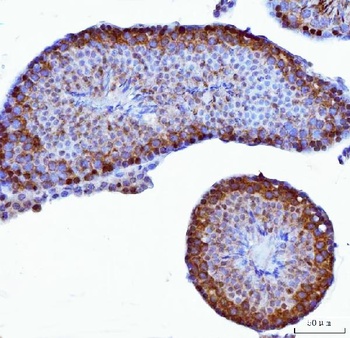
IHC analysis of YBX1 using anti-YBX1 antibody (orb76271). YBX1 was detected in a paraffin-embedded section of mouse testis tissue. Heat mediated antigen retrieval was performed in EDTA buffer (pH 8.0, epitope retrieval solution). The tissue section was blocked with 10% goat serum. The tissue section was then incubated with 2 µg/ml rabbit anti-YBX1 Antibody (orb76271) overnight at 4C. Peroxidase Conjugated Goat Anti-rabbit IgG was used as secondary antibody and incubated for 30 minutes at 37C. The tissue section was developed using HRP Conjugated Rabbit IgG Super Vision Assay Kit with DAB as the chromogen.
IHC analysis of YBX1 using anti-YBX1 antibody (orb76271). YBX1 was detected in a paraffin-embedded section of rat testis tissue. Heat mediated antigen retrieval was performed in EDTA buffer (pH 8.0, epitope retrieval solution). The tissue section was blocked with 10% goat serum. The tissue section was then incubated with 2 µg/ml rabbit anti-YBX1 Antibody (orb76271) overnight at 4C. Peroxidase Conjugated Goat Anti-rabbit IgG was used as secondary antibody and incubated for 30 minutes at 37C. The tissue section was developed using HRP Conjugated Rabbit IgG Super Vision Assay Kit with DAB as the chromogen.
Flow Cytometry analysis of HEL cells using anti-YBX1 antibody (orb76271). Overlay histogram showing HEL cells st
* Mehrwertsteuer und Versandkosten nicht enthalten. Irrtümer und Preisänderungen vorbehalten