E.coli-derived human PC4/SUB1 recombinant protein (Position: N62-L127).
Konjugation:
Unconjugated
Alternative Synonym:
Neuronal PAS domain-containing protein 2, Neuronal PAS2, Basic-helix-loop-helix-PAS protein MOP4, Class E basic helix-loop-helix protein 9, bHLHe9, Member of PAS protein 4, PAS domain-containing protein 4, NPAS2, BHLHE9, MOP4, PASD4
Anti-PC4/SUB1 Antibody. Tested in ELISA, IF, IHC, WB applications. This antibody reacts with Human, Mouse, Rat.
Klonalität:
Polyclonal
Konzentration:
Adding 0.2 ml of distilled water will yield a concentration of 500 µg/ml.
Activated RNA polymerase II transcriptional coactivator p15
Application Verdünnung:
Western blot, 0.1-0.25 µg/ml, Human, Mouse, Rat Immunohistochemistry(Paraffin-embedded Section), 2-5 µg/ml, Human Immunofluorescence, 5 µg/ml, Human ELISA, 0.1-0.5 µg/ml, -
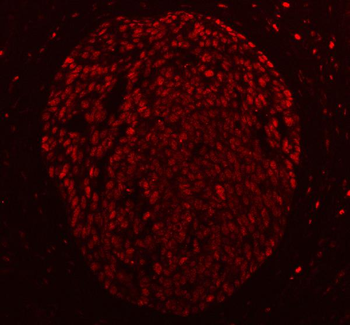
IF analysis of PC4/SUB1 using anti-PC4/SUB1 antibody. PC4/SUB1 was detected in a paraffin-embedded section of human cervical carcinoma tissue. Heat mediated antigen retrieval was performed in EDTA buffer (pH8.0, epitope retrieval solution). The tissue section was blocked with 10% goat serum. The tissue section was then incubated with 5 µg/mL rabbit anti-PC4/SUB1 Antibody overnight at 4C. Cy3 Conjugated Goat Anti-Rabbit IgG was used as secondary antibody at 1:500 dilution and incubated for 30 minutes at 37C. Visualize using a fluorescence microscope and filter sets appropriate for the label used.
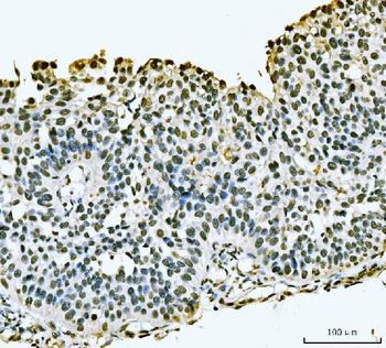
IHC analysis of PC4/SUB1 using anti-PC4/SUB1 antibody. PC4/SUB1 was detected in a paraffin-embedded section of human bladder epithelial carcinoma tissue. Heat mediated antigen retrieval was performed in EDTA buffer (pH8.0, epitope retrieval solution). The tissue section was blocked with 10% goat serum. The tissue section was then incubated with 2 µg/ml rabbit anti-PC4/SUB1 Antibody overnight at 4C. Biotinylated goat anti-rabbit IgG was used as secondary antibody and incubated for 30 minutes at 37C. The tissue section was developed using Strepavidin-Biotin-Complex (SABC) with DAB as the chromogen.
IHC analysis of PC4/SUB1 using anti-PC4/SUB1 antibody. PC4/SUB1 was detected in a paraffin-embedded section of human gastric signet ring cell carcinoma tissue. Heat mediated antigen retrieval was performed in EDTA buffer (pH8.0, epitope retrieval solution). The tissue section was blocked with 10% goat serum. The tissue section was then incubated with 2 µg/ml rabbit anti-PC4/SUB1 Antibody overnight at 4C. Biotinylated goat anti-rabbit IgG was used as secondary antibody and incubated for 30 minutes at 37C. The tissue section was developed using Strepavidin-Biotin-Complex (SABC) with DAB as the chromogen.
IHC analysis of PC4/SUB1 using anti-PC4/SUB1 antibody. PC4/SUB1 was detected in a paraffin-embedded section of human gastric signet ring cell carcinoma tissue. Heat mediated antigen retrieval was performed in EDTA buffer (pH8.0, epitope retrieval solution). The tissue section was blocked with 10% goat serum. The tissue section was then incubated with 2 µg/ml rabbit anti-PC4/SUB1 Antibody overnight at 4C. Biotinylated goat anti-rabbit IgG was used as secondary antibody and incubated for 30 minutes at 37C. The tissue section was developed using Strepavidin-Biotin-Complex (SABC) with DAB as the chromogen.
IHC analysis of PC4/SUB1 using anti-PC4/SUB1 antibody. PC4/SUB1 was detected in a paraffin-embedded section of human papillary carcinoma of the left breast tissue. Heat mediated antigen retrieval was performed in EDTA buffer (pH8.0, epitope retrieval solution). The tissue section was blocked with 10% goat serum. The tissue section was then incubated with 2 µg/ml rabbit anti-PC4/SUB1 Antibody overnight at 4C. Biotinylated goat anti-rabbit IgG was used as secondary antibody and incubated for 30 minutes at 37C. The tissue section was developed using Strepavidin-Biotin-Complex (SABC) with DAB as the chromogen.
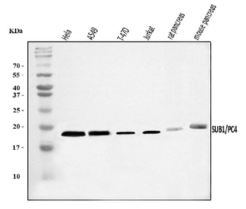
Western blot analysis of PC4/SUB1 using anti-PC4/SUB1 antibody. Electrophoresis was performed on a 5-20% SDS-PAGE gel at 70V (Stacking gel) / 90V (Resolving gel) for 2-3 hours. The sample well of each lane was loaded with 30 ug of sample under reducing conditions. Lane 1: human Hela whole cell lysates, Lane 2: human A549 whole cell lysates, Lane 3: human T-47D whole cell lysates, Lane 4: human Jurkat whole cell lysates, Lane 5: rat pancreas tissue lysates, Lane 6: mouse pancreas tissue lysates. After electrophoresis, proteins were transferred to a nitrocellulose membrane at 150 mA for 50-90 minutes. Blocked the membrane with 5% non-fat milk/TBS for 1.5 hour at RT. The membrane was incubated with rabbit anti-PC4/SUB1 antigen affinity purified polyclonal antibody at 0.25 µg/mL overnight at 4C, then washed with TBS-0.1% Tween 3 times with 5 minutes each and probed with a goat anti-rabbit IgG-HRP secondary antibody at a dilution of 1:500
* Mehrwertsteuer und Versandkosten nicht enthalten. Irrtümer und Preisänderungen vorbehalten