
Phospho-NPM1 (S125) Recombinant Monoclonal Antibody, Clone: [12B1], Unconjugated, Rabbit
Artikelnummer:
CSB-RA170404A0HU
- Bilder (5)
| Artikelname: | Phospho-NPM1 (S125) Recombinant Monoclonal Antibody, Clone: [12B1], Unconjugated, Rabbit |
| Artikelnummer: | CSB-RA170404A0HU |
| Hersteller Artikelnummer: | CSB-RA170404A0HU |
| Alternativnummer: | CSB-RA170404A0HU-100UL, CSB-RA170404A0HU-50UL |
| Hersteller: | Cusabio |
| Wirt: | Rabbit |
| Kategorie: | Antikörper |
| Applikation: | ELISA, FC, IF, IHC, WB |
| Spezies Reaktivität: | Human, Mouse |
| Konjugation: | Unconjugated |
| Alternative Synonym: | B23 antibody, MGC104254 antibody, NO38 antibody, NPM antibody, NPM_HUMAN antibody, NPM1 antibody, Nucleolar phosphoprotein B23 antibody, Nucleolar protein NO38 antibody, Nucleophosmin (nucleolar phosphoprotein B23 numatrin) antibody, Nucleophosmin antibody, nucleophosmin nucleoplasmin family member 1 antibody, Nucleophosmin/nucleoplasmin family member 1 antibody, Numatrin antibody, OTTHUMP00000161024 antibody, OTTHUMP00000161025 antibody, OTTHUMP00000223397 antibody, OTTHUMP00000223398 antibody |
| Klonalität: | Monoclonal |
| Klon-Bezeichnung: | [12B1] |
| UniProt: | P06748 |
| Puffer: | Rabbit IgG in 10mM phosphate buffered saline , pH 7.4, 150mM sodium chloride, 0.05% BSA, 0.02% sodium azide and 50% glycerol. |
| Reinheit: | Affinity-chromatography |
| Formulierung: | Liquid |
| Target-Kategorie: | NPM1 |
| Antibody Type: | Recombinant Antibody |
| Application Verdünnung: | Recommended dilution: WB:1:500-1:5000, IHC:1:50-1:200, IF:1:50-1:200, FC:1:50-1:200 |
|
|
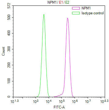
Overlay Peak curve showing jurkat cells stained with CSB-RA170404A0HU (red line) at 1:100. The cells were fixed in 4% formaldehyde and permeated by 0.2% TritonX-100 for10min. Then 10% normal goat serum to block non-specific protein-protein interactions followed by the antibody (1ug/1*106cells) for 45min at 4°C. The secondary antibody used was FITC-conjugated goat anti-rabbit IgG (H+L) at 1/200 dilution for 35min at 4°C.Control antibody (green line) was Rabbit IgG (1ug/1*106cells) used under the same conditions. Acquisition of >10, 000 events was performed. |
|
|
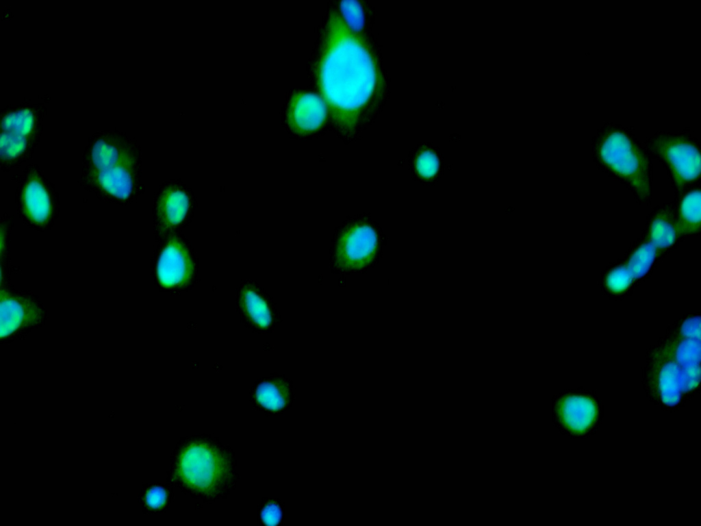
Immunofluorescence staining of HepG2 cell with CSB-RA170404A0HU at 1:50 , counter-stained with DAPI. The cells were fixed in 4% formaldehyde, permeabilized using 0.2% Triton X-100 and blocked in 10% normal Goat Serum. The cells were then incubated with the antibody overnight at 4C. The secondary antibody was Alexa Fluor 488-congugated AffiniPure Goat Anti-Rabbit IgG(H+L). |
|
|
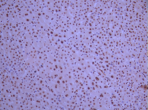
IHC image of CSB-RA170404A0HU diluted at 1:100 and staining in paraffin-embedded human liver tissue performed on a Leica BondTM system. After dewaxing and hydration, antigen retrieval was mediated by high pressure in a citrate buffer (pH 6.0). Section was blocked with 10% normal goat serum 30min at RT. Then primary antibody (1% BSA) was incubated at 4C overnight. The primary is detected by a Goat anti-rabbit polymer IgG labeled by HRP and visualized using 0.05% DAB. |
|
|
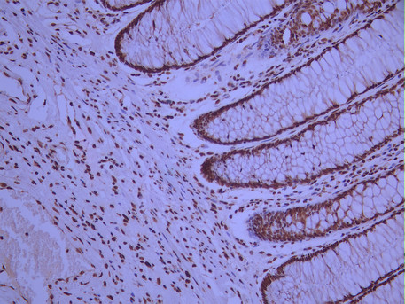
IHC image of CSB-RA170404A0HU diluted at 1:100 and staining in paraffin-embedded human colorectal cancer performed on a Leica BondTM system. After dewaxing and hydration, antigen retrieval was mediated by high pressure in a citrate buffer (pH 6.0). Section was blocked with 10% normal goat serum 30min at RT. Then primary antibody (1% BSA) was incubated at 4C overnight. The primary is detected by a Goat anti-rabbit polymer IgG labeled by HRP and visualized using 0.05% DAB. |
|
|
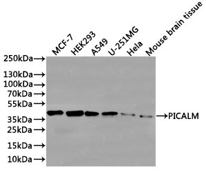
Western Blot Positive WB detected in: MCF7 whole cell lysate(30µg), HEK293 whole cell lysate(30µg), A549 whole cell lysate(30µg), U251 whole cell lysate(30µg), Hela whole cell lysate(30µg), Mouse brain tissue lysate(30µg) All lanes: p-Nucleophosmin (S125) antibody at 1:1000 Secondary Goat polyclonal to rabbit IgG at 1/40000 dilution Predicted band size: 33 kDa Observed band size: 38 kDa Exposure time:2min |
Produktgarantie und fachkundiger Support
