
CTBP2 Recombinant Monoclonal Antibody, Clone: [8D8], Unconjugated, Rabbit
Artikelnummer:
CSB-RA257842A0HU
- Bilder (5)
| Artikelname: | CTBP2 Recombinant Monoclonal Antibody, Clone: [8D8], Unconjugated, Rabbit |
| Artikelnummer: | CSB-RA257842A0HU |
| Hersteller Artikelnummer: | CSB-RA257842A0HU |
| Alternativnummer: | CSB-RA257842A0HU-100UL, CSB-RA257842A0HU-50UL |
| Hersteller: | Cusabio |
| Wirt: | Rabbit |
| Kategorie: | Antikörper |
| Applikation: | ELISA, FC, IF, IHC, WB |
| Spezies Reaktivität: | Human, Mouse, Rat |
| Konjugation: | Unconjugated |
| Alternative Synonym: | C terminal binding protein 2 antibody, C-terminal-binding protein 2 antibody, CtBP2 antibody, CTBP2_HUMAN antibody, ribeye antibody |
| Klonalität: | Monoclonal |
| Klon-Bezeichnung: | [8D8] |
| UniProt: | P56545 |
| Puffer: | Rabbit IgG in 10mM phosphate buffered saline , pH 7.4, 150mM sodium chloride, 0.05% BSA, 0.02% sodium azide and 50% glycerol. |
| Reinheit: | Affinity-chromatography |
| Formulierung: | Liquid |
| Target-Kategorie: | CTBP2 |
| Antibody Type: | Recombinant Antibody |
| Application Verdünnung: | Recommended dilution: WB:1:500-1:5000, IHC:1:50-1:200, IF:1:50-1:200, FC:1:50-1:200 |
|
|
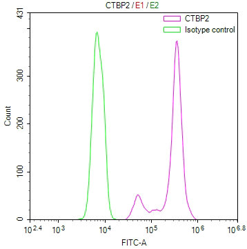
Overlay Peak curve showing Hela cells stained with CSB-RA257842A0HU (red line) at 1:100. The cells were fixed in 4% formaldehyde and permeated by 0.2% TritonX-100 for10min. Then 10% normal goat serum to block non-specific protein-protein interactions followed by the antibody (1ug/1*106cells) for 45min at 4°C. The secondary antibody used was FITC-conjugated goat anti-rabbit IgG (H+L) at 1/200 dilution for 35min at 4°C.Control antibody (green line) was Rabbit IgG (1ug/1*106cells) used under the same conditions. Acquisition of >10, 000 events was performed. |
|
|
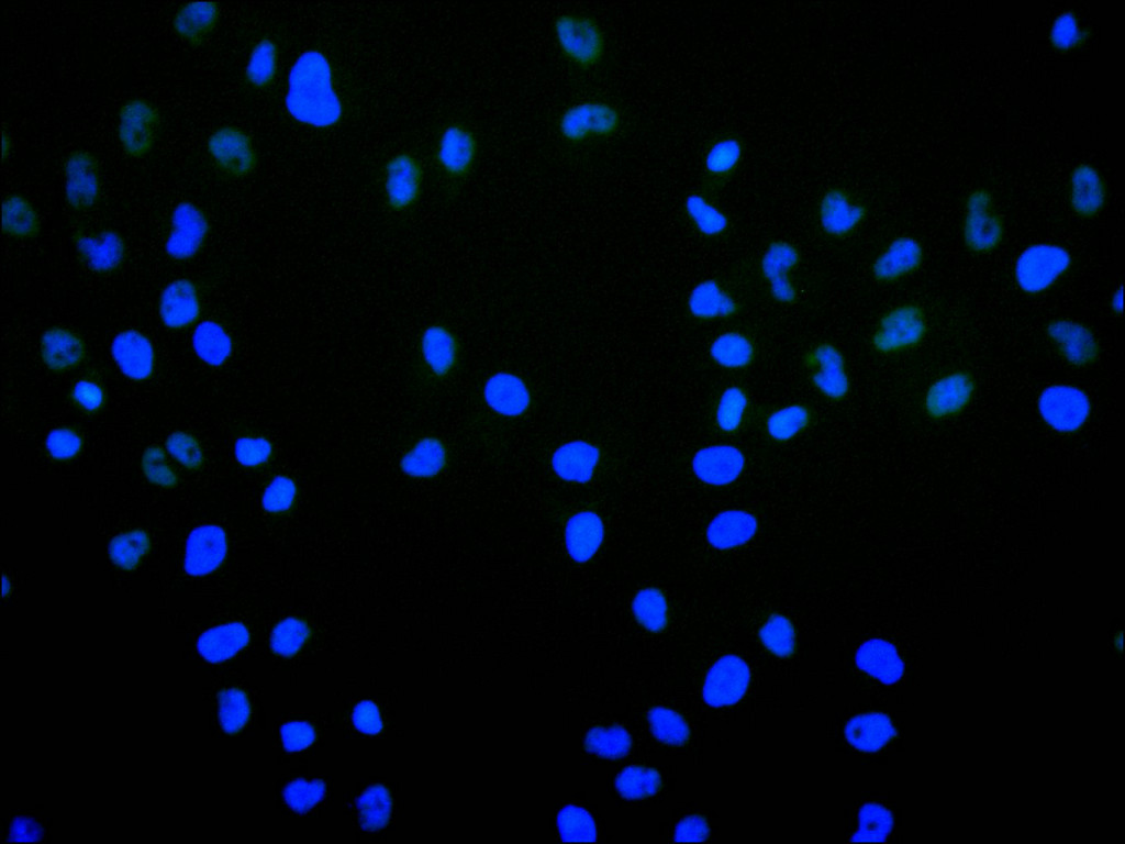
Immunofluorescence staining of HeLa cell with CSB-RA257842A0HU at 1:50 , counter-stained with DAPI. The cells were fixed in 4% formaldehyde, permeabilized using 0.2% Triton X-100 and blocked in 10% normal Goat Serum. The cells were then incubated with the antibody overnight at 4C. The secondary antibody was Alexa Fluor 488-congugated AffiniPure Goat Anti-Rabbit IgG(H+L). |
|
|
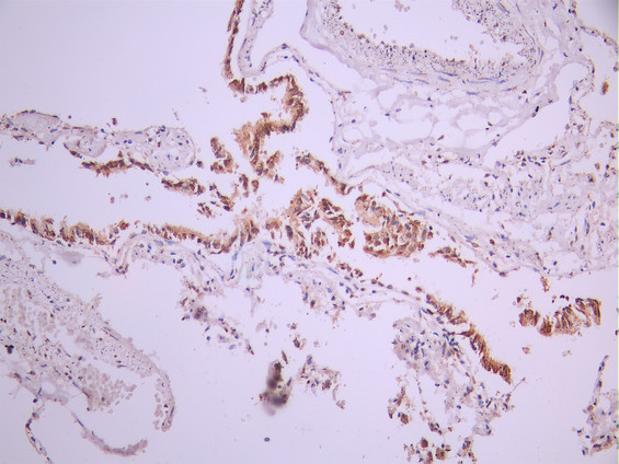
IHC image of CSB-RA257842A0HU diluted at 1:100 and staining in paraffin-embedded human lung tissue performed on a Leica BondTM system. After dewaxing and hydration, antigen retrieval was mediated by high pressure in a citrate buffer (pH 6.0). Section was blocked with 10% normal goat serum 30min at RT. Then primary antibody (1% BSA) was incubated at 4C overnight. The primary is detected by a Goat anti-rabbit polymer IgG labeled by HRP and visualized using 0.05% DAB. |
|
|
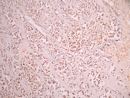
IHC image of CSB-RA257842A0HU diluted at 1:100 and staining in paraffin-embedded human breast cancer performed on a Leica BondTM system. After dewaxing and hydration, antigen retrieval was mediated by high pressure in a citrate buffer (pH 6.0). Section was blocked with 10% normal goat serum 30min at RT. Then primary antibody (1% BSA) was incubated at 4C overnight. The primary is detected by a Goat anti-rabbit polymer IgG labeled by HRP and visualized using 0.05% DAB. |
|
|
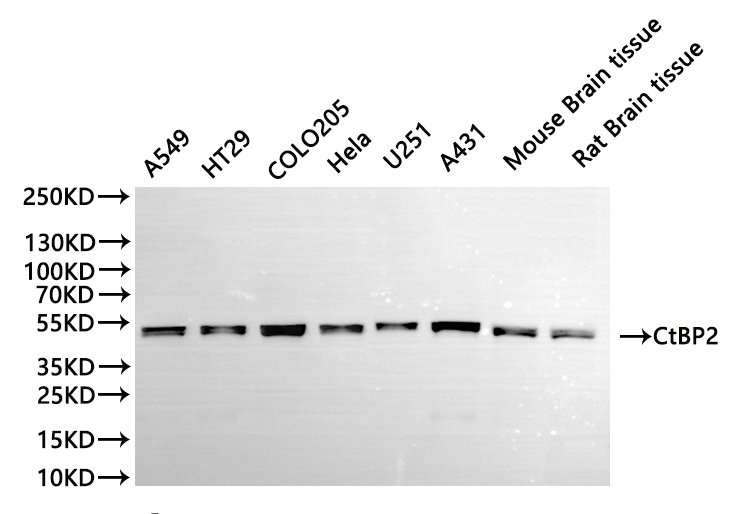
Western Blot , Positive WB detected in:A549 whole cell lysate(30µg), HT29 whole cell lysate(30µg), COLO205 whole cell lysate(30µg), HELA whole cell lysate(30µg), U251 whole cell lysate(30µg), A431 whole cell lysate(30µg), Mouse brain tissue lysate(30µg), Rat brain lysate(30µg) All lanes: CtBP2 antibody at 1:1000 Secondary Goat polyclonal to rabbit IgG at 1/40000 dilution Predicted band size: 49 kDa Observed band size: 49 kDa Exposure time:10s |
Produktgarantie und fachkundiger Support
