
SCP2 Recombinant Monoclonal Antibody, Clone: [11E6], Unconjugated, Rabbit
Artikelnummer:
CSB-RA286943A0HU
- Bilder (5)
| Artikelname: | SCP2 Recombinant Monoclonal Antibody, Clone: [11E6], Unconjugated, Rabbit |
| Artikelnummer: | CSB-RA286943A0HU |
| Hersteller Artikelnummer: | CSB-RA286943A0HU |
| Alternativnummer: | CSB-RA286943A0HU-100UL, CSB-RA286943A0HU-50UL |
| Hersteller: | Cusabio |
| Wirt: | Rabbit |
| Kategorie: | Antikörper |
| Applikation: | ELISA, FC, IF, IHC, WB |
| Spezies Reaktivität: | Human, Mouse |
| Konjugation: | Unconjugated |
| Alternative Synonym: | DKFZp686C12188 antibody, DKFZp686D11188 antibody, NLTP antibody, NLTP_HUMAN antibody, Non-specific lipid-transfer protein antibody, Nonspecific lipid transfer protein antibody, NSL TP antibody, NSL-TP antibody, OTTHUMP00000010488 antibody, OTTHUMP00000231766 antibody, OTTHUMP00000231767 antibody, OTTHUMP00000231768 antibody, OTTHUMP00000231769 antibody, OTTHUMP00000231770 antibody, OTTHUMP00000231772 antibody, OTTHUMP00000231773 antibody, OTTHUMP00000231774 antibody, OTTHUMP00000231776 antibody, OTTHUMP00000234662 antibody, Propanoyl CoA C acyltransferase antibody, Propanoyl-CoA C-acyltransferase antibody, SCP 2 antibody, SCP chi antibody, SCP X antibody, SCP-2 antibody, SCP-chi antibody, SCP-X antibody, SCP2 antibody, SCPchi antibody, SCPX antibody, Sterol carrier protein 2 antibody, Sterol carrier protein X antibody |
| Klonalität: | Monoclonal |
| Klon-Bezeichnung: | [11E6] |
| UniProt: | P22307 |
| Puffer: | Rabbit IgG in 10mM phosphate buffered saline , pH 7.4, 150mM sodium chloride, 0.05% BSA, 0.02% sodium azide and 50% glycerol. |
| Reinheit: | Affinity-chromatography |
| Formulierung: | Liquid |
| Target-Kategorie: | SCP2 |
| Antibody Type: | Recombinant Antibody |
| Application Verdünnung: | Recommended dilution: WB:1:500-1:5000, IHC:1:50-1:200, IF:1:50-1:200, FC:1:50-1:200 |
|
|
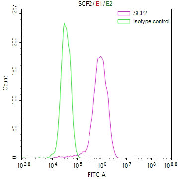
Overlay Peak curve showing MCF-7 cells stained with CSB-RA286943A0HU (red line) at 1:100. The cells were fixed in 4% formaldehyde and permeated by 0.2% TritonX-100 for10min. Then 10% normal goat serum to block non-specific protein-protein interactions followed by the antibody (1ug/1*106cells) for 45min at 4°C. The secondary antibody used was FITC-conjugated goat anti-rabbit IgG (H+L) at 1/200 dilution for 35min at 4°C.Control antibody (green line) was Rabbit IgG (1ug/1*106cells) used under the same conditions. Acquisition of >10, 000 events was performed. |
|
|
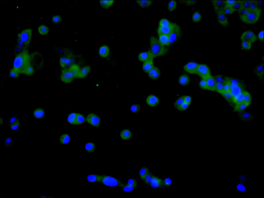
Immunofluorescence staining of HepG2 cell with CSB-RA286943A0HU at 1:50 , counter-stained with DAPI. The cells were fixed in 4% formaldehyde, permeabilized using 0.2% Triton X-100 and blocked in 10% normal Goat Serum. The cells were then incubated with the antibody overnight at 4C. The secondary antibody was Alexa Fluor 488-congugated AffiniPure Goat Anti-Rabbit IgG(H+L). |
|
|
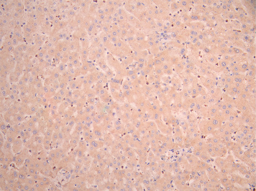
IHC image of CSB-RA286943A0HU diluted at 1:100 and staining in paraffin-embedded human liver tissue performed on a Leica BondTM system. After dewaxing and hydration, antigen retrieval was mediated by high pressure in a citrate buffer (pH 6.0). Section was blocked with 10% normal goat serum 30min at RT. Then primary antibody (1% BSA) was incubated at 4C overnight. The primary is detected by a Goat anti-rabbit polymer IgG labeled by HRP and visualized using 0.05% DAB. |
|
|
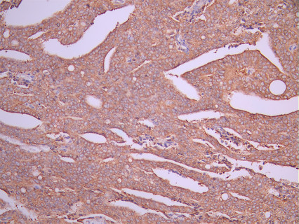
IHC image of CSB-RA286943A0HU diluted at 1:100 and staining in paraffin-embedded human prostate cancer performed on a Leica BondTM system. After dewaxing and hydration, antigen retrieval was mediated by high pressure in a citrate buffer (pH 6.0). Section was blocked with 10% normal goat serum 30min at RT. Then primary antibody (1% BSA) was incubated at 4C overnight. The primary is detected by a Goat anti-rabbit polymer IgG labeled by HRP and visualized using 0.05% DAB. |
|
|
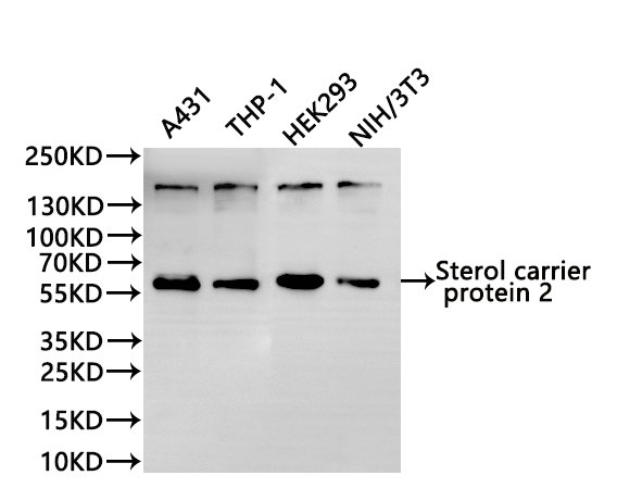
Western Blot Positive WB detected in: A431 whole cell lysate(30µg), THP-1 whole cell lysate(30µg), HEK293 whole cell lysate(30µg), NIH/3T3 whole cell lysate(30µg) All lanes:Sterol carrier protein 2 antibody at 1:1000 Secondary Goat polyclonal to rabbit IgG at 1/40000 dilution Predicted band size: 59 kDa Observed band size: 59 kDa Exposure time:1min |
Produktgarantie und fachkundiger Support
