
CASP8 Recombinant Monoclonal Antibody, Clone: [6A9], Unconjugated, Rabbit
Artikelnummer:
CSB-RA549188A0HU
- Bilder (5)
| Artikelname: | CASP8 Recombinant Monoclonal Antibody, Clone: [6A9], Unconjugated, Rabbit |
| Artikelnummer: | CSB-RA549188A0HU |
| Hersteller Artikelnummer: | CSB-RA549188A0HU |
| Alternativnummer: | CSB-RA549188A0HU-100UL, CSB-RA549188A0HU-50UL |
| Hersteller: | Cusabio |
| Wirt: | Rabbit |
| Kategorie: | Antikörper |
| Applikation: | ELISA, FC, IF, IHC, WB |
| Spezies Reaktivität: | Human |
| Konjugation: | Unconjugated |
| Alternative Synonym: | ALPS2B antibody, Amyotrophic lateral sclerosis 2 chromosomal region candidate gene 12 protein antibody, Apoptosis related cysteine peptidase antibody, Apoptotic cysteine protease antibody, Apoptotic protease Mch-5 antibody, Apoptotic protease Mch5 antibody, CAP 4 antibody, CAP4 antibody, CASP-8 antibody, CASP8 antibody, CASP8_HUMAN antibody, Caspase 8 antibody, Caspase 8 apoptosis related cysteine peptidase antibody, Caspase IIX antibody, Caspase-8 subunit p10 antibody, caspase8 antibody, CED 3 antibody, FADD Homologous ICE/CED3 Like Protease antibody, FADD Like ICE antibody, FADD-homologous ICE/CED-3-like protease antibody, FADD-like ICE antibody, FLICE antibody, FLJ17672 antibody, ICE-like apoptotic protease 5 antibody, MACH alpha 1/2/3 protein antibody, MACH antibody, MACH beta 1/2/3/4 protein antibody, MACH5 antibody, MCH 5 antibody, MCH5 antibody, MGC78473 antibody, MORT1 associated ced 3 homolog antibody, MORT1 associated CED3 homolog antibody, MORT1-associated CED-3 homolog antibody, OTTHUMP00000163717 antibody, OTTHUMP00000163720 antibody, OTTHUMP00000163724 antibody, OTTHUMP00000163725 antibody, OTTHUMP00000165062 antibody, OTTHUMP00000165063 antibody, OTTHUMP00000165064 antibody, OTTHUMP00000206552 antibody, OTTHUMP00000206582 antibody |
| Klonalität: | Monoclonal |
| Klon-Bezeichnung: | [6A9] |
| UniProt: | Q14790 |
| Puffer: | Rabbit IgG in 10mM phosphate buffered saline , pH 7.4, 150mM sodium chloride, 0.05% BSA, 0.02% sodium azide and 50% glycerol. |
| Reinheit: | Affinity-chromatography |
| Formulierung: | Liquid |
| Target-Kategorie: | CASP8 |
| Antibody Type: | Recombinant Antibody |
| Application Verdünnung: | Recommended dilution: WB:1:500-1:5000, IHC:1:50-1:200, IF:1:50-1:200, FC:1:50-1:200 |
|
|
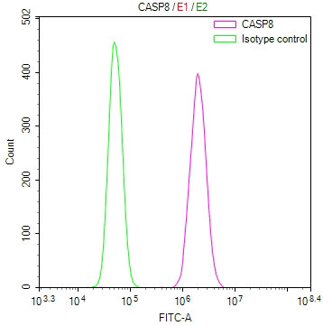
Overlay Peak curve showing 786-O cells stained with CSB-RA549188A0HU (red line) at 1:100. The cells were fixed in 4% formaldehyde and permeated by 0.2% TritonX-100 for10min. Then 10% normal goat serum to block non-specific protein-protein interactions followed by the antibody (1ug/1*106cells) for 45min at 4°C. The secondary antibody used was FITC-conjugated goat anti-rabbit IgG (H+L) at 1/200 dilution for 35min at 4°C.Control antibody (green line) was Rabbit IgG (1ug/1*106cells) used under the same conditions. Acquisition of >10, 000 events was performed. |
|
|
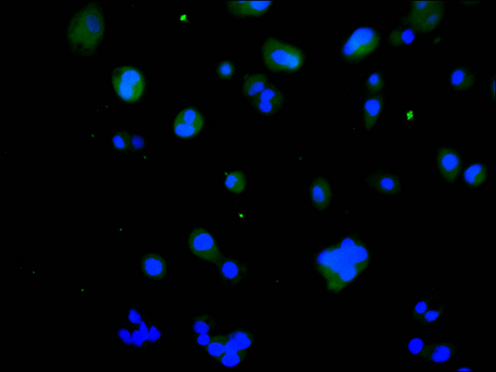
Immunofluorescence staining of HepG2 cell with CSB-RA549188A0HU at 1:50 , counter-stained with DAPI. The cells were fixed in 4% formaldehyde, permeabilized using 0.2% Triton X-100 and blocked in 10% normal Goat Serum. The cells were then incubated with the antibody overnight at 4C. The secondary antibody was Alexa Fluor 488-congugated AffiniPure Goat Anti-Rabbit IgG(H+L). |
|
|
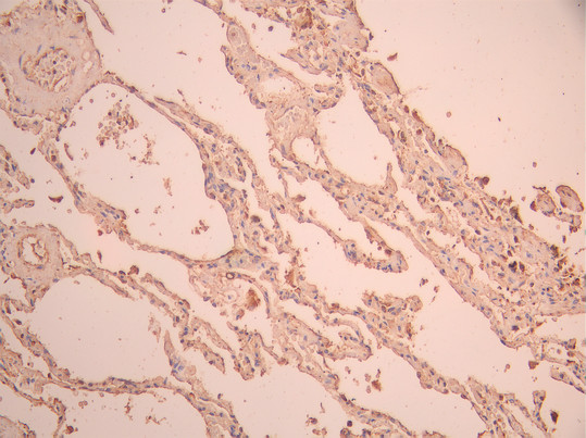
IHC image of CSB-RA549188A0HU diluted at 1:100 and staining in paraffin-embedded human lung tissue performed on a Leica BondTM system. After dewaxing and hydration, antigen retrieval was mediated by high pressure in a citrate buffer (pH 6.0). Section was blocked with 10% normal goat serum 30min at RT. Then primary antibody (1% BSA) was incubated at 4C overnight. The primary is detected by a Goat anti-rabbit polymer IgG labeled by HRP and visualized using 0.05% DAB. |
|
|
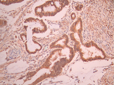
IHC image of CSB-RA549188A0HU diluted at 1:100 and staining in paraffin-embedded human gastric cancer performed on a Leica BondTM system. After dewaxing and hydration, antigen retrieval was mediated by high pressure in a citrate buffer (pH 6.0). Section was blocked with 10% normal goat serum 30min at RT. Then primary antibody (1% BSA) was incubated at 4C overnight. The primary is detected by a Goat anti-rabbit polymer IgG labeled by HRP and visualized using 0.05% DAB. |
|
|
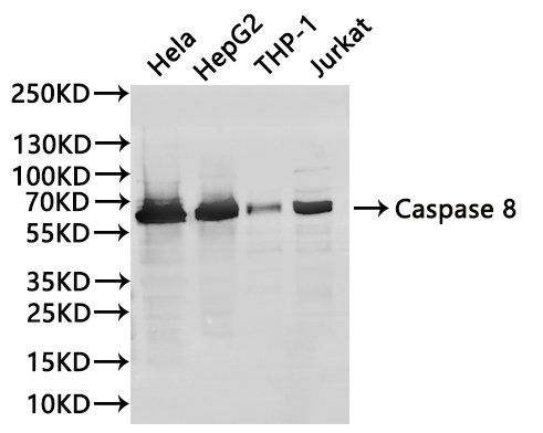
Western Blot , Positive WB detected in:Hela whole cell lysate(30µg), HepG2 whole cell lysate(30µg), THP-1 whole cell lysate(30µg), Jurkat whole cell lysate(30µg) All lanes: Caspase 8 antibody at 1:1000 Secondary Goat polyclonal to rabbit IgG at 1/40000 dilution Predicted band size: 55 kDa Observed band size: 62 kDa Exposure time:40s |
Produktgarantie und fachkundiger Support
