
ACAA2 Recombinant Monoclonal Antibody, Clone: [12F4], Unconjugated, Rabbit
Artikelnummer:
CSB-RA978277A0HU
- Bilder (5)
| Artikelname: | ACAA2 Recombinant Monoclonal Antibody, Clone: [12F4], Unconjugated, Rabbit |
| Artikelnummer: | CSB-RA978277A0HU |
| Hersteller Artikelnummer: | CSB-RA978277A0HU |
| Alternativnummer: | CSB-RA978277A0HU-100UL, CSB-RA978277A0HU-50UL |
| Hersteller: | Cusabio |
| Wirt: | Rabbit |
| Kategorie: | Antikörper |
| Applikation: | ELISA, IF, IHC, WB |
| Spezies Reaktivität: | Human, Mouse |
| Konjugation: | Unconjugated |
| Alternative Synonym: | 3-ketoacyl-CoA thiolase, mitochondrial (EC 2.3.1.16) (Acetyl-CoA acyltransferase) (Beta-ketothiolase) (Mitochondrial 3-oxoacyl-CoA thiolase) (T1) , ACAA2 |
| Klonalität: | Monoclonal |
| Klon-Bezeichnung: | [12F4] |
| UniProt: | P42765 |
| Puffer: | Rabbit IgG in 10mM phosphate buffered saline , pH 7.4, 150mM sodium chloride, 0.05% BSA, 0.02% sodium azide and 50% glycerol. |
| Reinheit: | Affinity-chromatography |
| Formulierung: | Liquid |
| Target-Kategorie: | ACAA2 |
| Antibody Type: | Recombinant Antibody |
| Application Verdünnung: | Recommended dilution: WB:1:500-1:2000, IHC:1:50-1:200, IF:1:50-1:200 |
|
|
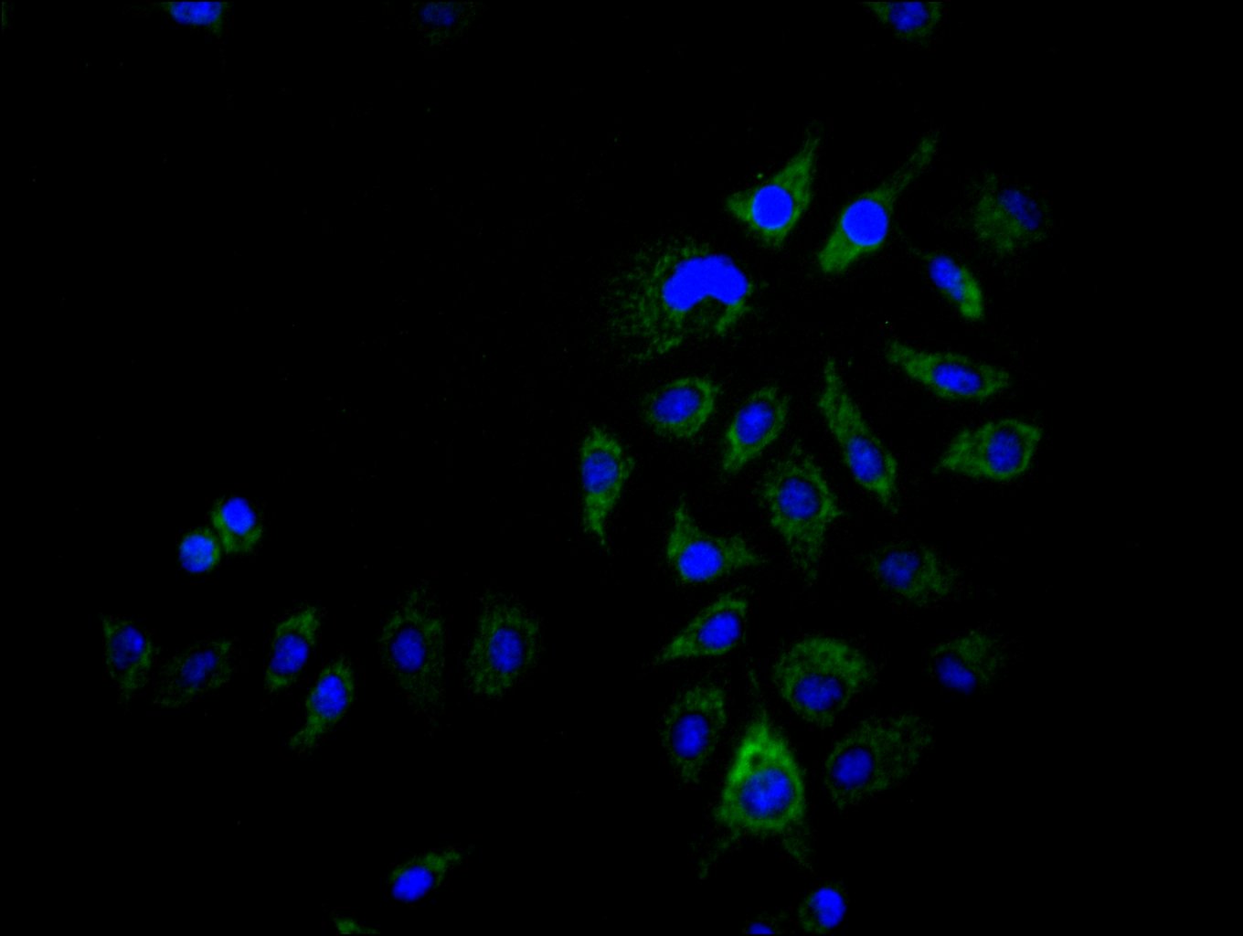
Immunofluorescence staining of Hela cell with CSB-RA978277A0HU at 1:12, counter-stained with DAPI. The cells were fixed in 4% formaldehyde and blocked in 10% normal Goat Serum. The cells were then incubated with the antibody overnight at 4C. The secondary antibody was Alexa Fluor 488-congugated AffiniPure Goat Anti-Rabbit IgG(H+L) . |
|
|
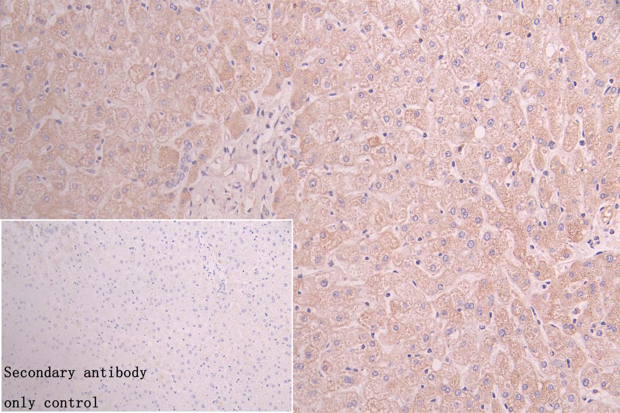
IHC image of CSB-RA978277A0HU diluted at 1:150 and staining in paraffin-embedded human liver tissue performed on a Leica BondTM system. After dewaxing and hydration, antigen retrieval was mediated by high pressure in a citrate buffer (pH 6.0) . Section was blocked with 10% normal goat serum 30min at RT. Then primary antibody (1% BSA) was incubated at 4C overnight. The primary is detected by a Goat anti-rabbit polymer IgG labeled by HRP and visualized using 0.05% DAB. Secondary antibody only control: uses 1% BSA instead of primary antibody |
|
|
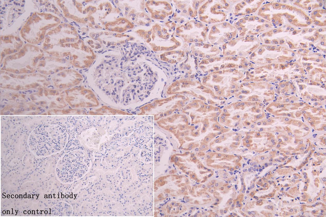
IHC image of CSB-RA978277A0HU diluted at 1:150 and staining in paraffin-embedded human kidney tissue performed on a Leica BondTM system. After dewaxing and hydration, antigen retrieval was mediated by high pressure in a citrate buffer (pH 6.0) . Section was blocked with 10% normal goat serum 30min at RT. Then primary antibody (1% BSA) was incubated at 4C overnight. The primary is detected by a Goat anti-rabbit polymer IgG labeled by HRP and visualized using 0.05% DAB. Secondary antibody only control: uses 1% BSA instead of primary antibody |
|
|
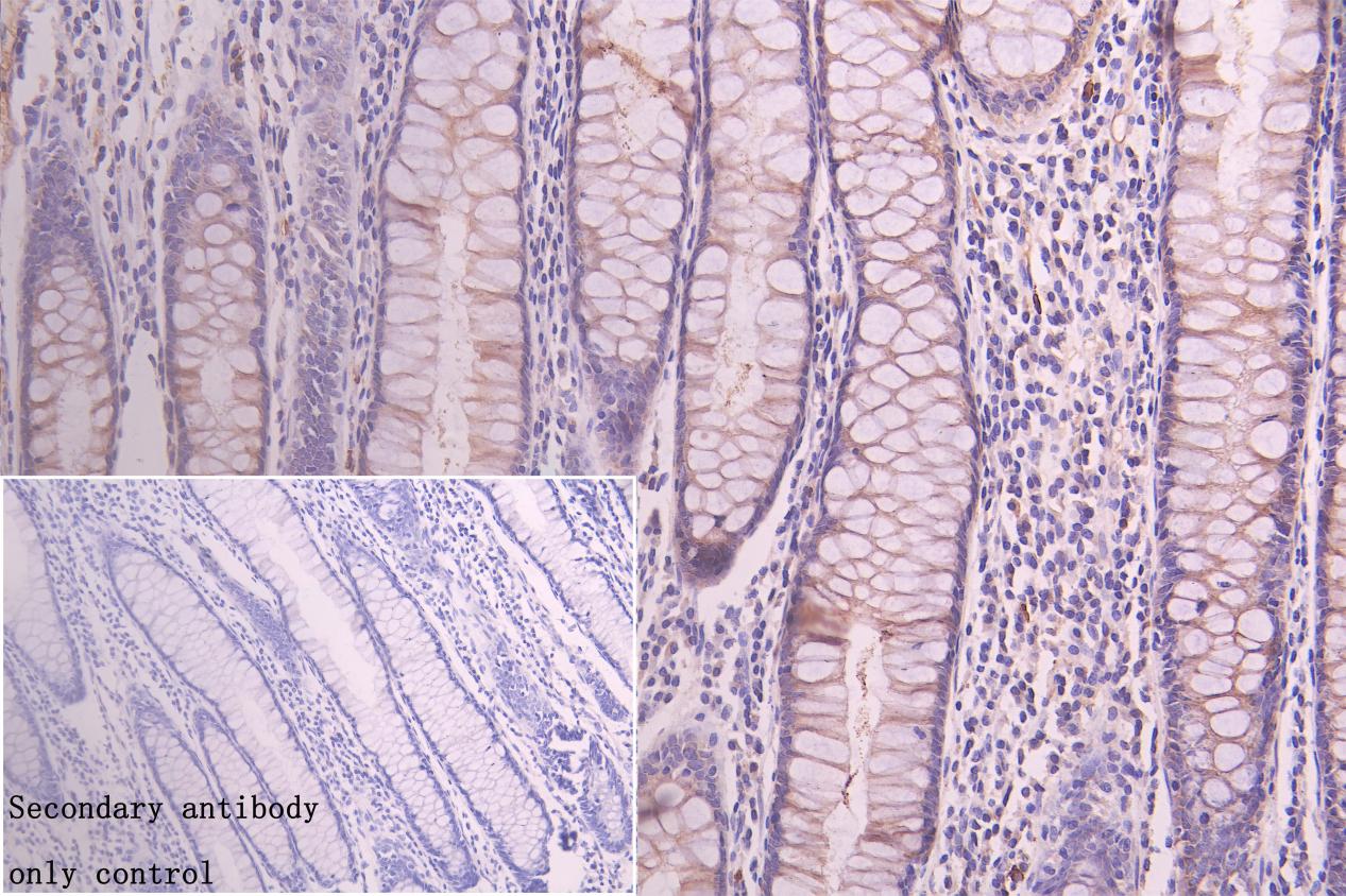
IHC image of CSB-RA978277A0HU diluted at 1:150 and staining in paraffin-embedded human colorectal cancer performed on a Leica BondTM system. After dewaxing and hydration, antigen retrieval was mediated by high pressure in a citrate buffer (pH 6.0) . Section was blocked with 10% normal goat serum 30min at RT. Then primary antibody (1% BSA) was incubated at 4C overnight. The primary is detected by a Goat anti-rabbit polymer IgG labeled by HRP and visualized using 0.05% DAB. Secondary antibody only control: uses 1% BSA instead of primary antibody |
|
|
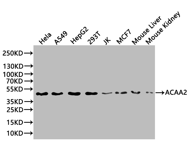
Western Blot Positive WB detected in: Hela whole cell lysate(20µg) , A549 whole cell lysate(20µg) , HepG2 whole cell lysate(20µg) , 293T whole cell lysate(20µg) , MCF7 whole cell lysate(20µg) , JK whole cell lysate(20µg) , Mouse Liver tissue lysate(20µg) , Mouse Kidney tissue lysate(20µg) All lanes: ACAA2 antibody at 1:1000 Secondary Goat polyclonal to rabbit IgG at 1/50000 dilution Predicted band size: 42 kDa Observed band size: 42 kDa Exposure time: 10s |
Produktgarantie und fachkundiger Support
