Synthesized peptide derived from Endophilin I . at AA range: 30-110
Alternative Synonym:
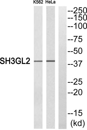
SH3GL2, CNSA2, SH3D2A, Endophilin-A1, EEN-B1, Endophilin-1, SH3 domain protein 2A, SH3 domain-containing GRB2-like protein 2
domain:An N-terminal amphipathic helix, the BAR domain and a second amphipathic helix inserted into helix 1 of the BAR domain (N-BAR domain) induce membrane curvature and bind curved membranes. The BAR domain dimer forms a rigid crescent shaped bundle of helices with the pair of second amphipathic helices protruding towards the membrane-binding surface.,function:Implicated in synaptic vesicle endocytosis. May recruit other proteins to membranes with high curvature.,miscellaneous:HeLa cells expressing the N-BAR domain of SH3GL2 show tubulation of the plasma membrane. The N-BAR domain binds liposomes and induces formation of tubules from liposomes. The N-terminal amphipathic helix is required for liposome binding. The second amphipathic helix enhances liposome tubulation.,similarity:Belongs to the endophilin family.,similarity:Contains 1 BAR domain.,similarity:Contains 1 SH3 domain.,subcellular location:Concentrated in presynaptic nerve terminals in neurons.,subunit:Monomer, in cytoplasm. Homodimer, when associated with membranes (By similarity). Interacts with SYNJ1 and DNM1. Interacts with MAP4K3, the interaction appears to regulate MAP4K3-mediated JNK activation. Interacts with PDCD6IP.,tissue specificity:Brain, mostly in frontal cortex. Expressed at high level in fetal cerebellum.,