This whole rabbit serum was prepared by repeated immunizations with a synthetic peptide corresponding to the C-terminal region of murine Dab1 at amino acids 400-555.
0.02 M Potassium Phosphate, 0.15 M Sodium Chloride, pH 7.2
Formulierung:
Liquid (sterile filtered)
Target-Kategorie:
Mouse
Antibody Type:
Primary Antibody
Application Verdünnung:
ELISA: 1:10,000 - 1:50,000, IHC: 1:5,000, IF Microscopy: User Optimized, IP: 1 µl per 500 µg, WB: 1:5,000
Anwendungsbeschreibung:
Anti-Dab1 antibody has been tested by western blot and immunohistochemistry and is suitable for the detection of Dab1 by immunoprecipitation. Specific conditions for reactivity should be optimized by the end user. Expect a band at 80 kDa corresponding to
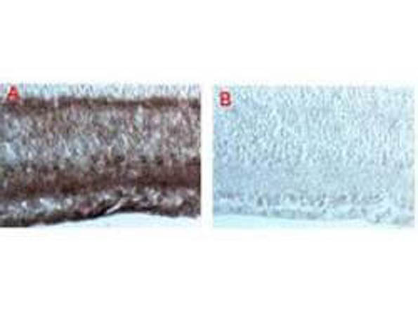
Immunohistochemical staining after cryofixation and sectioning of mouse brain tissue using (A) a 1:5,000 dilution of anti-Dab-1 and (B) 1:5,000 dilution of pre-immune serum followed by processing with HRP Goat anti-Rabbit IgG [H&L] and chromogenic substrate.
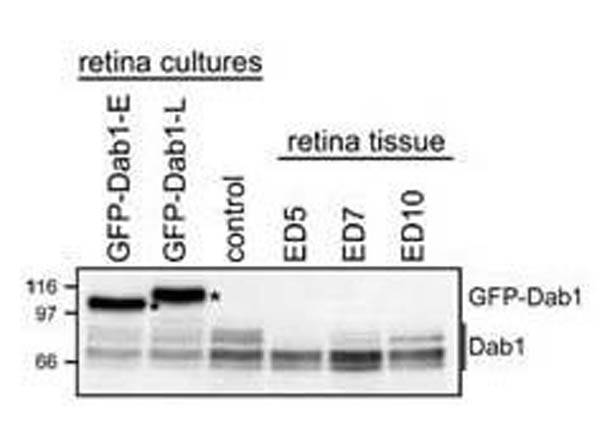
Analysis of GFP-Dab1 and endogenous Dab1 levels in transfected retinal cells and retinal tissue. Western blot analysis of whole cell lysates prepared from primary retinal cultures transfected with GFP-Dab1-E (lane 1), -L (lane 2), control untransfected retinal cultures (lane 3), and retinal tissue at ED5 (lane 4), ED7 (lane 5) and ED10 (lane 6). Proteins were electrophoresed through an SDS-8% polyacrylamide gel and transferred to nitrocellulose. The membrane was immuno-stained with Rockland Immunochemicals anti-Dab1 antibody at 1:5000, which recognizes both GFP-Dab1 (indicated by asterisks) and endogenous forms of Dab1 (indicated by a line). See Katval et al (2007) for additional details.
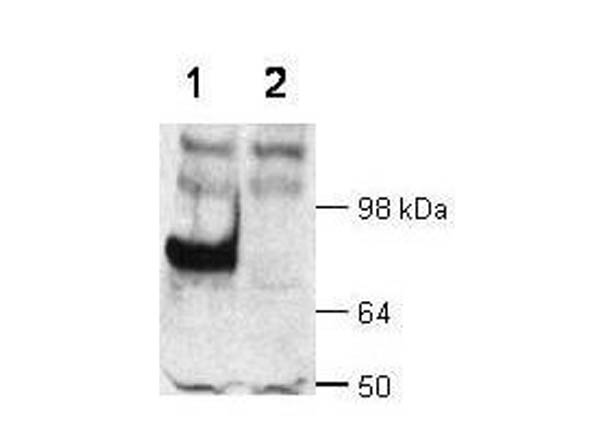
Rockland Immunochemicals anti-Dab1 is shown to detect Dab1 present in wt mouse brain extracts (lane 1). No staining is noted in similar extracts from a dab knock-out mouse (lane 2). Detection of an 80 kDa band (arrowhead) occurs using a 1:5,000 dilution of the antibody in 1% milk in TTBS for 1 h at room temperature followed by a 1:5,000 dilution of HRP Goat-a-Rabbit with ECL visualization. Film exposure was ~1. Other detection systems will yield similar results.
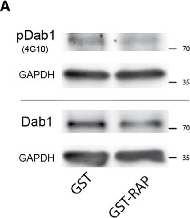
Coapplication of GST-RAP does not prevent the E2-induced distal dendritic HCN1 enrichment in vitro. A, Western blots showing phosphorylated Dab1 (pDab1, upper bands, detected by anti-phosphotyrosine antibody 4G10) and total Dab1 (lower bands) in ''paired slice cultures that were treated for 24 h (DIV10-11) with either GST-RAP or GST (10 µg/ml each) and were exposed to Reelin-conditioned medium for 30 min before harvesting. GAPDH was used for loading control. B, Quantitative analysis of Dab1 and pDab1 levels (relative to GAPDH) revealed that Reelin-induced pDab1 was reduced in the GST-RAP-treated slices, while total Dab1 was not significantly different compared to the GST-treated controls. Thus, pDab1/Dab1 was reduced to 72% 6% of control levels after 24-h GST-RAP treatment (p < 0.01, n = 15). C, D, E2 (+GST)-treatment (pink) of cultures for 6 d (DIV5-11) caused a significant HCN1 accumulation in segment 5 (C) and a significantly increased slope (D) compared with controls (GST, black) that was not efficiently reduced, if GST-RAP (10 µg/ml) was coapplied (orange, n = 17, each group). E-G, Representative photographs from a culture ''triple, of which one culture served as a vehicle (GST)-treated control (E), while the others were treated with either E2 + GST (F) or E2 + GST-RAP (G). Note that HCN1 is accumulated at the hippocampal fissure (asterisks) at all conditions, but if E2 was present (F, G), less HCN1 immunosignal is visible in stratum radiatum (sr, arrows), indicating (relative) HCN1 enrichment in stratum lacunosum-moleculare (slm). Scale bars: 80 µm (E-G). Dashed lines demarcate the border of stratum pyramidale (sp). ml, molecular layer. Figure provided by CiteAb. Source: Eneuro, PMID: 30406178.
* Mehrwertsteuer und Versandkosten nicht enthalten. Irrtümer und Preisänderungen vorbehalten