
TNF alpha Antibody, Rabbit, Polyclonal
Artikelnummer:
ROC-109-401-306S
- Bilder (5)

| Artikelname: | TNF alpha Antibody, Rabbit, Polyclonal |
| Artikelnummer: | ROC-109-401-306S |
| Hersteller Artikelnummer: | 109-401-306S |
| Alternativnummer: | ROC-109-401-306S |
| Hersteller: | Rockland Immunochemicals |
| Wirt: | Rabbit |
| Kategorie: | Antikörper |
| Applikation: | IF, IHC, WB |
| Spezies Reaktivität: | Human, Primate |
| Immunogen: | The whole rabbit serum was prepared by repeated immunizations with recombinant human TNFa produced in E.coli. |
| Konjugation: | Unconjugated |
| Alternative Synonym: | APC1 antibody, Cachectin antibody, DIF antibody, Differentiation inducing factor antibody, Macrophage cytotoxic factor antibody, MCF antibody, Necrosin antibody, Tumour Necrosis Factor Alpha antibody, rabbit anti-Tumor Necrosis Factor Alpha Antibody, rabbit anti-TNF Alpha Antibody, Tumor necrosis factor, TNF-alpha, Tumor necrosis factor ligand superfamily member 2, TNF-a, Tumor necrosis factor membrane form, N-terminal fragment, NTF, Intracellular domain 1, ICD1, Intracellular domain 2, ICD2, C-domain 1, C-domain 2, Tumor necrosis factor, soluble form, TNF, TNFA, TNFSF2 |
| Application Verdünnung: | ELISA: 1:1,000 - 1:5,000, IHC: 1:100 - 1:500, IF Microscopy: 1:100, WB: 1:500 - 1:2,000 |
| Anwendungsbeschreibung: | Anti-Human TNFalpha has been tested for use in immunohistochemistry, immunofluorescence, and immunoblotting. It recognizes the 17,000 MW TNFalpha. Reactivity in other immunoassays is unknown. |
|
|
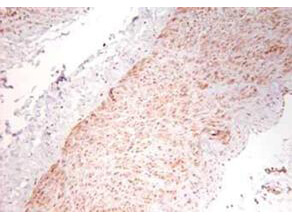
Immunohistochemistry using Rocklands polyclonal TNFa antibody showing staining of formalin/PFA-fixed paraffin-embedded sections of human artery tissue sections. Sections were fixed in formaldehyde and subjected to heat mediated antigen retrieval in citrate buffer (pH 6.0). Slides were blocked for ten minutes with 1.5% serum. Primary antibody was diluted 1:100 and incubated with samples for 24 hours at 4C. HRP-conjugated goat anti-rabbit antibody was used as the secondary antibody. |
|
|
|
|
|
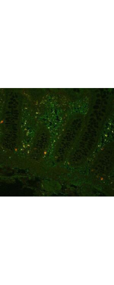
Fluorescent immunohistochemistry showing staining of human colon by Rocklands anti-TNF alpha (formalin/PFA-fixed paraffin-embedded sections). Samples were formaldehyde-fixed, then blocked in 10% serum for 2 hours at 20C. The primary antibody was diluted 1:100 and incubated with the sample for 2 hours at 20C. Alexa Fluor 680 goat polyclonal secondary antibody was used diluted 1:5000. |
|
|
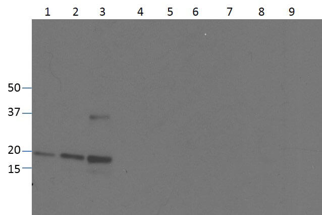
Western blot of Anti-Human TNF-a (RABBIT) Antibody. Lane 1: 250ng human recombinant TNF. Lane 2: 500ng human recombinant TNF. Lane 3: 1000ng human recombinant TNF. Membrane blocked in Blotto (p/n B501-0500) for 30 min RT. Primary antibody: Rb-a-TNF alpha added at 1:1000 in overnight at 4C, Gt-a-Rb HRP (p/n 611-103-122) added at 1:20,000 in 5% Blotto for 30 min RT. (5 second exposure) |
|
|
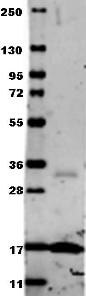
Western blot using Rocklands Anti-Human TNF-a (RABBIT) Antibody. Membrane blocked in 1% BSA-TBS-T 30 min RT, Rb-a-TNF alpha added at 1:1000 in 1% BSA-TBS-T o/n 4C, DyLight 649 Gt-a-Rb (p/n 611-143-122) added at 1:20,000 in (p/n MB-070) 30 min RT. |
Produktgarantie und fachkundiger Support
