Anti-Keratin Antibody has been tested in ELISA, immunohistochemistry, immunofluorescence, immunoblotting and immunoprecipitation. For a positive control use skin, colon carcinoma and squamous granulocyte carcinoma cells.
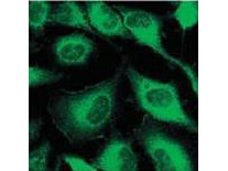
Immunofluorescence using ROCKLAND Immunochemicals Mouse Anti-Keratin antibody. Confocal slices of HeLa cells are between 0.5 and 0.6 µm where the image is taken near the bottom of the cell. Use FITC conjugated Goat-a-Mouse IgG [H&L] (p/n 610-102-121) at 1:2,000 dilution for detection.
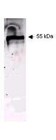
Western blot using ROCKLAND Immunochemicals Mouse Anti-Keratin antibody. This antibody recognizes a single 56kDa band corresponding to human keratin as confirmed by the position of molecular weight markers (not shown). Approximately 100ng of keratin from human epidermis was applied under reducing conditions to a pre-cast 4-20% iGel from Gradipore Inc. A 1:400 dilution of Mab anti-Keratin was used for 2h followed by detection using a 1:5,000 dilution of IRDye(TM)800 conjugated Goat-a-Mouse IgG [H&L] (p/n 610-132-121) and visualization using the Odyssey Infrared Imaging System developed by LI-COR. Other detection systems will yield similar results. IRDye is a trademark of LI-COR, Inc.
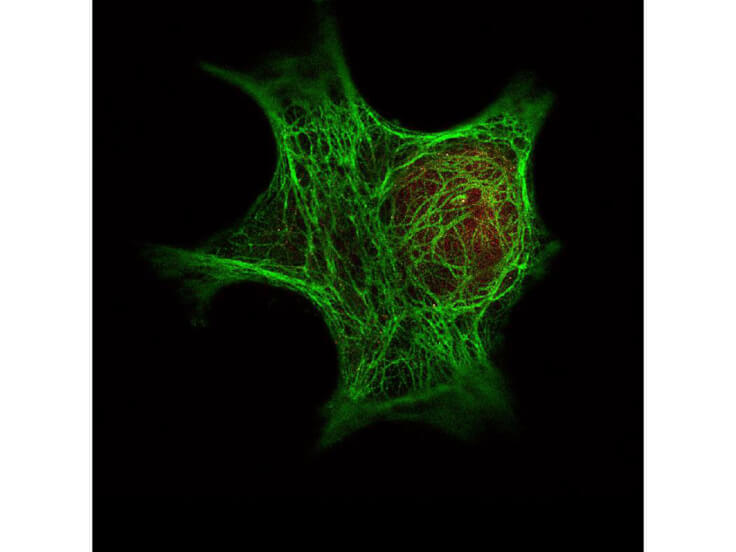
Immunofluorescence Microscopy of Rockland Immunochemicals Anti-Keratin antibody (p/n 200-301-390) was used with Rocklands DyLight(TM) 488 goat anti-mouse (p/n 610-141-121) [shown in green] to detect Keratin by Immunofluorescence. In the same experiment, Rocklands polyclonal Anti-HDAC-1 antibody (p/n 600-401-879) was used with Atto425 Anti-Rabbit IgG (p/n 611-151-122) [shown in red] to detect HDAC-1. Data was collected on a STED-CW TCS-SP5 Confocal system (Leica Microsystems) equipped with a DFC 350FX camera allowing sequential acquisition in wide-field, confocal and STED CW imaging modes and provided courtesy of: Myriam Gastard, PhD, personal communication, Leica Microsystems, Inc. USA
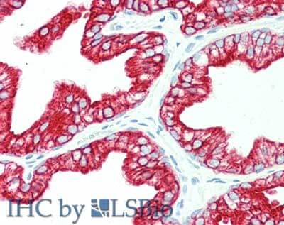
Immunohistochemistry of Mouse anti-Keratin antibody. Tissue: human prostate. Fixation: formalin fixed paraffin embedded. Antigen retrieval: not required. Primary antibody: anti-Keratin antibody at 10 µg/mL for 1 h at RT. Secondary antibody: Peroxidase mouse secondary antibody at 1:10,000 for 45 min at RT. Staining: Keratin as precipitated red signal with hematoxylin purple nuclear counterstain.
* Mehrwertsteuer und Versandkosten nicht enthalten. Irrtümer und Preisänderungen vorbehalten