0.1 M Tris Chloride, 0.5 M Sodium Chloride, pH 8.0
Formulierung:
Liquid (sterile filtered)
Target-Kategorie:
Rabbit
Antibody Type:
Primary Antibody
Application Verdünnung:
ELISA: User Optimized, IHC: User Optimized, IF Microscopy: 1:100, IP: User Optimized, WB: 1:1,000
Anwendungsbeschreibung:
Anti-GAPDH (Mouse) has been tested in ELISA and western blot. This product is suitable in IHC and IF. Specific conditions should be optimized by the end user. Expect a band of ~38kDa in size corresponding to Glyceraldehyde 3-Phosphate Dehydrogenase prote
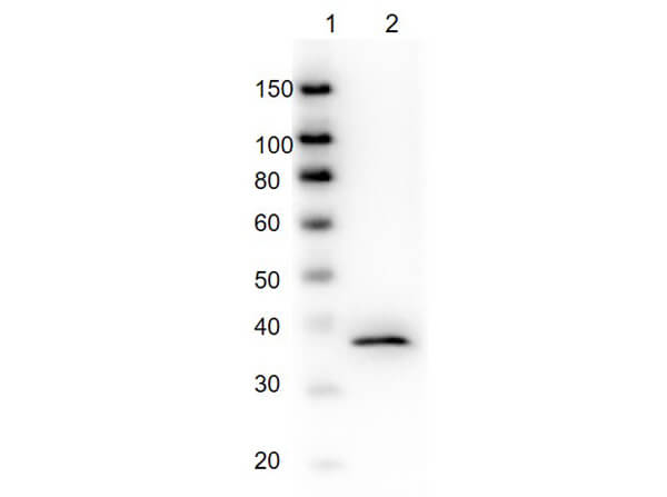
Western Blot of Mouse Anti-GAPDH Antibody. Lane 1: Molecular Weight. Lane 2: Glyceraldehyde-3-Phosphate-Dehydrogenase. Primary Antibody: Mouse Anti-GAPDH Antibody at 1µg/mL. Secondary Antibody: Anti-Mouse IgG HRP. Block: BlockOut Buffer (p/n MB-073). Predicted MW: ~38kDa.
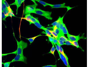
Immunofluorescence Microscopy of Mouse Anti-Glyceraldehyde 3-Phosphate Dehydrogenase Antibody. Tissue: Human neuroblastoma SH-SY5Y cells. Fixation: 0.5% PFA. Antigen retrieval: not required. Primary antibody: GAPDH antibody at 10 µg/mL for 1 h at RT. Secondary antibody: Fluorescein mouse secondary antibody at 1:10,000 for 45 min at RT. Localization: GAPDH is cytoplasmic. Staining: Anti-GAPDH (green), chicken antibody to neurofilament NF-H (red) and DNA (blue).
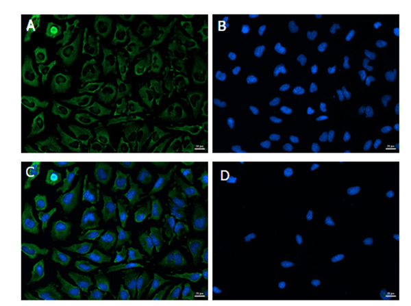
Immunofluorescence Microscopy of Mouse Anti-Glyceraldehyde 3-Phosphate Dehydrogenase Antibody. Cells: HeLa cells. Fixation: Ice Cold Methanol. Permeabilization: Ice Cold Methanol. Primary antibody: GAPDH antibody at 4µg/mL (1:250) overnight at 4C. Secondary antibody: Goat anti-Mouse DyLight(TM)488 (p/n 610-141-121) at 1:1,000 for 1hr at RT. Localization: GAPDH is cytoplasmic. Staining: (A) Anti-GAPDH (green), (B) Hoechst Nuclear stain, (C) merge, (D) secondary only and Hoechst Nuclear stain. Magnification Objective: 20x.
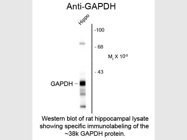
Western Blot of Mouse anti-Glyceraldehyde-3-Phosphate Dehydrogenase antibody. Lane 1: rat hippocampal lysate. Lane 2: none. Load: 10 µg per lane. Primary antibody: GAPDH antibody at 1:400 for overnight at 4C. Secondary antibody: IRDye800(TM) mouse secondary antibody at 1:10,000 for 45 min at RT. Block: 5% BLOTTO overnight at 4C. Predicted/Observed size: ~38kDa/~38kDa for GAPDH protein. Other band(s): splice variants and isoforms.
* Mehrwertsteuer und Versandkosten nicht enthalten. Irrtümer und Preisänderungen vorbehalten