This antibody was produced in mesothelin-deficient mice by immunizations with plasmid cDNA encoding human MSLN full length protein followed by a single boost of a recombinant human mesothelin-Fc fusion protein.
This antibody has been tested for use in immunohistochemistry, Flow cytometry, and western blotting. Specific conditions for reactivity should be optimized by the end user. Expect a band approximately 40 kDa in size corresponding to mature mesothelin by
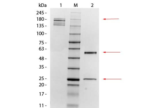
SDS-PAGE of Mouse anti-Mesothelin Monoclonal Antibody. Lane 1: Non-Reduced Mouse anti-Mesothelin Monoclonal Antibody. Lane M: 3 µL OPAL Pre-stained Marker (p/n MB-210-0500). Lane 2: Reduced Mouse anti-Mesothelin Monoclonal Antibody. Load: 1 µg per lane. Predicted/Observed size: Non-reduced at 160 kDa, Reduced at 55, 25 kDa.
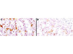
Immunohistochemistry using Rocklands anti-mesothelin antibody to react with two epitopes on mesothelin in PEFF human mesothelioma tissue sections treated by antigen retrieval methods. Anti-mesothelin primary antibodies were used at 10 µg/mL to label these sections as follows: C, MAb MB, and D, MAb MN followed by goat anti-mouse IgG conjugated to horseradish peroxidase at 25 µg/mL in 1% BSA/PBS for 30 minutes. (magnification, *200, bar, 50 µm). Reprinted with permission from Clin.Cancer Res. 11(16):5840-6.
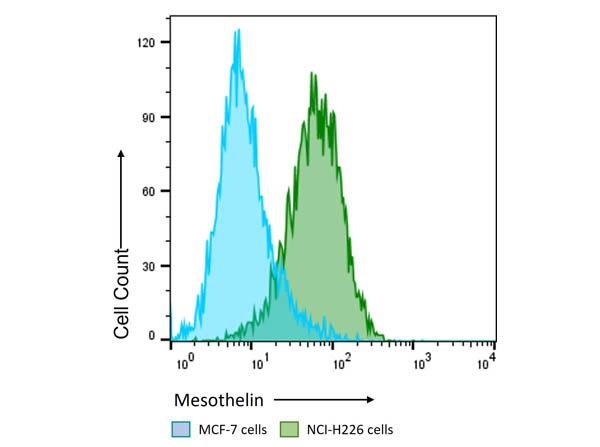
Flow Cytometry Results of Anti-Mesothelin (MOUSE) Monoclonal Antibody. The green histogram shows NCI-H226 cells and blue histogram shows MCF-7 cells. Both cell lines are stained with a 1:200 dilution Anti-Mesothelin (MOUSE) Monoclonal Antibody. The secondary antibody use was Anti-Mouse IgG (H&L) (GOAT) Antibody DyLight(TM) 488 Conjugated (p/n 610-141-002, lot43322) at the 1:400 dilution.
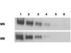
Western blotting using Rocklands anti-mesothelin antibody. Load: Mesothelin-Fc (lane 1, 100 ng, lane 2, 25 ng, lane 3, 6 ng, lane 4, 2 ng, and lane 5, 0.4 ng) and CD25-Fc (lane 6, 50 ng) Primary antibody: anti-mesothelin at 1mg/ml. Secondary Antibody: ALP goat anti-mouse IgG and BCIP/NBT substrate. Reprinted with permission from Clin.Cancer Res. 11(16):5840-6.
* Mehrwertsteuer und Versandkosten nicht enthalten. Irrtümer und Preisänderungen vorbehalten