This monoclonal antibody was produced by repeated immunizations with a synthetic peptide corresponding to a region near the carboxy terminus of human TRPC6 protein.
Konjugation:
Unconjugated
Alternative Synonym:
mouse anti-TRPC6 Antibody, TRPC 6, TRP6, short transient receptor potential channel 6 and transient receptor potential cation channel subfamily C member 6
Anti-TRPC6 monoclonal antibody has been tested by ELISA, immunohistochemistry and western blotting. Expect a band approximately 30 kDa in size corresponding to the cytoplasmic domain of TRPC6 protein by western blotting in the appropriate cell lysate or
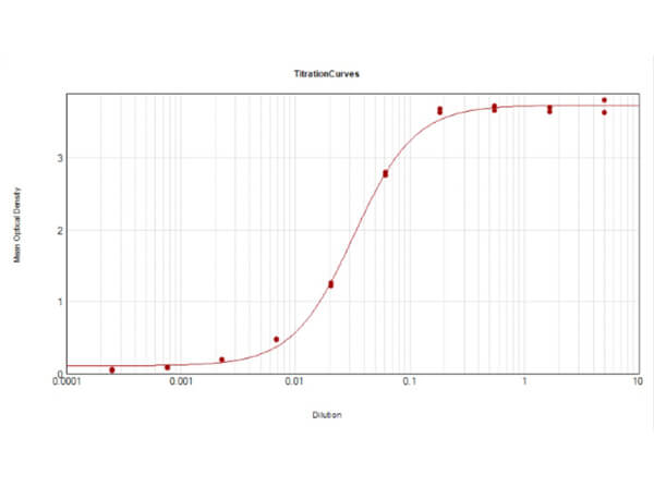
ELISA Results of Mouse Anti-TRPC6 Antibody. Each well was coated in duplicate with 0.1µg of conjugate. The working dilution is 1:31,000. The starting dilution of antibody was 5µg/ml and the X-axis represents the Log10 of a 3-fold dilution. This titration is a 4-parameter curve fit where the IC50 is defined as the titer of the antibody. Assay performed using HRP conjugation Stabilizer (p/n MB-076), Rabbit Anti-Mouse IgG HRP conjugated (p/n 610-403-C46) and TMB substrate (p/n TMBE-1000).
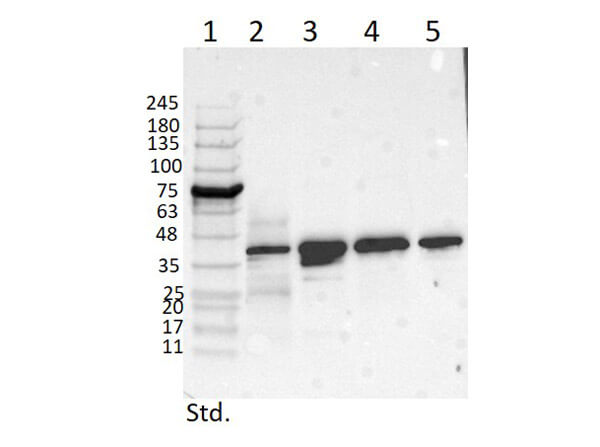
Western Blot of Mouse Anti-TRPC6 Antibody. Lane 1: Opal Prestained Molecular Weight Marker (p/n MB-210-0500). Lane 2: Mouse Pancreas Tissue Lysate (p/n W10-000-T023) [10µg]. Lane 3: MCF-7 Whole Cell Lysate (p/n W09-000-360) [10µg]. Lane 4: A431 Whole Cell Lysate (p/n W09-000-361) [10µg]. Lane 5: Jurkat Whole Cell Lysate (p/n W09-001-370) [10µg]. Primary Antibody: Anti-TRPC6 at 1µg/mL overnight at 2-8C. Secondary Antibody: Rabbit Anti-Mouse IgG Peroxidase (p/n 610-403-C46) 1:40000 for 30mins at RT. Blocking Buffer: BlockOut Buffer (p/n MB-073) for 30mins at RT. Predicted MW: ~30kDa. Observed MW: ~40kDa. Exposure: 5sec.
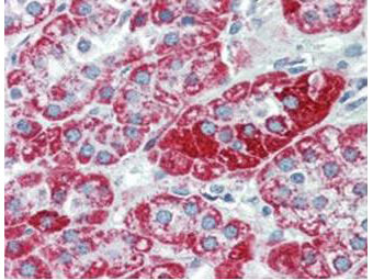
Immunohistochemistry using Rocklands anti-TRPC6 monoclonal antibody shows detection of TRPC6 in human adrenal (cortex) tissue (40X). The antibody was used a dilution to 2.5 µg/mL. The image shows strong staining with minimal background staining. Tissue was formalin fixed and paraffin embedded. No pre-treatment of sample was required. The image shows the localization of antibody as the precipitated red signal, with a hematoxylin purple nuclear counterstain. Personal communication, Andrew Elston, Lifespan Biosciences, Seattle, WA.
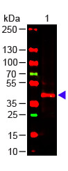
Western Blot of Mouse anti-TRPC6 Antibody. Lane 1: Mouse Kidney WCL (p/n W10-000-T017). Load: 10 µg per lane. Primary antibody: TRPC6 Antibody at 1:1000 for overnight at 4C. Secondary antibody: donkey anti-mouse DyLight(TM) 649 (p/n 610-743-002) at 1:20,000 for 30 min at RT. Block: MB-070 for 30 min at RT.
* Mehrwertsteuer und Versandkosten nicht enthalten. Irrtümer und Preisänderungen vorbehalten