Anti-PD-1 Antibody has been tested for use in ELISA, Western Blotting, Immunohistochemistry and Immunofluorescence. Specific conditions for reactivity should be optimized by the end user. Expect a band at approximately 32 kDa in Western Blots of specific
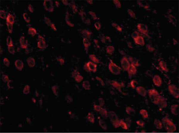
Immunofluorescence Microscopy of Mouse anti-PD-1 antibody. Tissue: mouse brain. Primary antibody: PD-1 antibody at 20 µg/mL. Secondary antibody: Peroxidase mouse secondary antibody at 1:20,000. Localization:PD-1 is located on the membrane. Staining: PD-1 as red fluorescent signal.
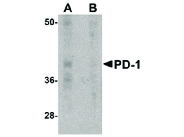
Western Blot of Mouse anti-PD-1 antibody. Lane A: A-20 cell lysate in absence of blocking recombinant protein. Lane B: A-20 cell lysate in the presence of blocking recombinant protein. Primary antibody: PD-1 antibody at 1 µg/mL overnight at 4°C. Secondary antibody: Mouse HRP secondary antibody. Block: 5% BLOTTO. Predicted/Observed size: 31 kDa, 39 kDa for PD-1.
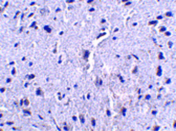
Immunohistochemistry of Mouse anti-PD-1 antibody. Tissue: mouse brain. Primary antibody: PD-1 antibody at 2.5 µg/mL. Secondary antibody: Peroxidase mouse secondary antibody. Localization: PD-1 is located on the membrane.
Immunofluorescence of PD-1. Tissue: human spleen tissue. Primary Antibody: PD-1 antibody at 20 µg/ml. Staining: PD-1 Antibody [green], DAPI staining [blue].
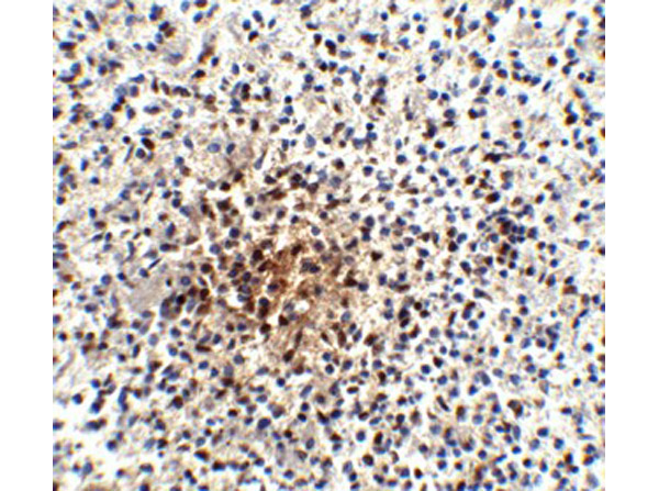
Immunohistochemistry of PD-1. Tissue: human spleen tissue. Primary Antibody: PD-1 antibody at 25 µg/ml.
* Mehrwertsteuer und Versandkosten nicht enthalten. Irrtümer und Preisänderungen vorbehalten