Anti-AKT1 Antibody is tested in ELISA, Flow Cytometry, and western blotting. This antibody is suitable in immunohistochemistry. Expect a band approximately 56 kDa in size corresponding to AKT1 protein by western blotting in the appropriate cell lysate or
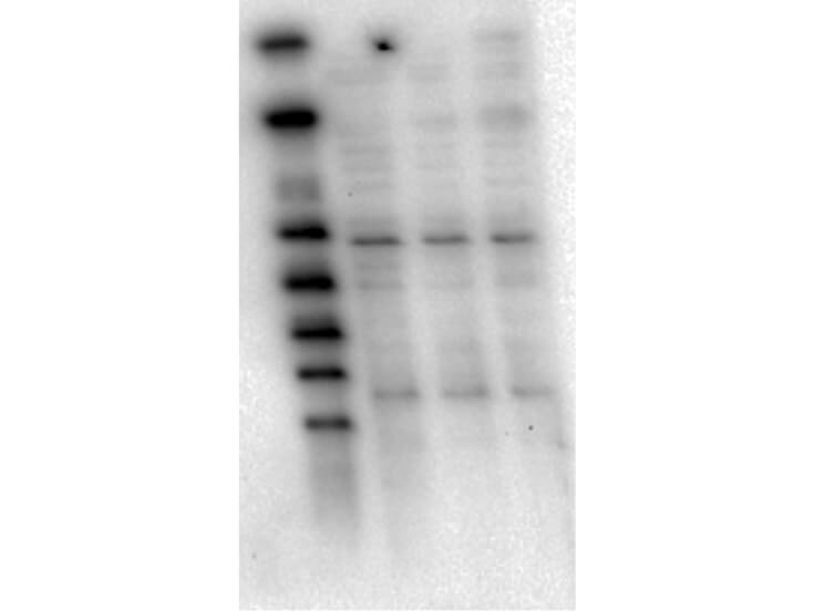
Western Blot of Mouse Anti-AKT1 antibody. Lane 1: Molecular Weight Marker. Lane 2: LnCap lysate (p/n W09-001-GJ9). Lane 3: Jurkat lysate (p/n W09-001-370). Lane 4: MDA-MB 468 lysate (p/n W09-001-GG9). Load: 5 µg per lane. Primary antibody: AKT1 antibody at 1:1000 for overnight at 4C. Secondary antibody: Mouse secondary antibody at 1:20,000 for 45 min at RT. Block: 5% BLOTTO overnight at 4C. Predicted/Observed size: 56 kDa for AKT1.
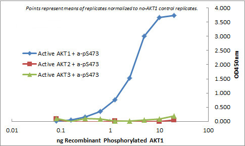
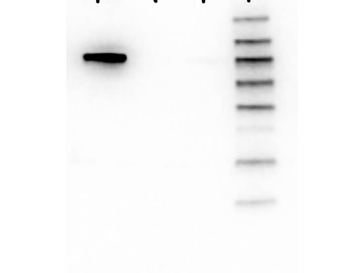
Western Blot of Mouse anti-AKT1 antibody. Lane 1: GST-AKT1. Lane 2: GST-AKT2. Lane 3: GST-AKT3. Lane 4: Molecular Weight Marker. Load: 25 ng per lane. Primary antibody: AKT1 antibody at 1:1000 for overnight at 4C. Secondary antibody: Mouse secondary antibody at 1:40,000 for 30 min at RT. Block: 5% BLOTTO overnight at 4C. Predicted/Observed size: 78 kDa for AKT1. Other band(s): none.
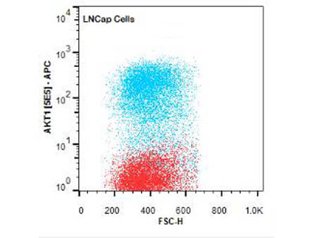
Flow Cytometry of Mouse anti-AKT1 antibody. Cells: LNCap Cells. Stimulation: none. Primary antibody: Allophycocyanin AKT1 antibody at 1.0 µg/mL for 20 min at 4C.
* Mehrwertsteuer und Versandkosten nicht enthalten. Irrtümer und Preisänderungen vorbehalten