Anti-5-mC Antibody was produced in mice by repeated immunization with 5-methyl Cytidine.
Konjugation:
Unconjugated
Alternative Synonym:
5-methylcytidine, 5-methyl Cytidine
Klonalität:
Monoclonal
Konzentration:
2.4 mg/ml by UV absorbance at 280 nm
Isotyp:
IgG1
Puffer:
0.01 M Sodium Phosphate, 0.25 M Sodium Chloride, pH 7.2
Formulierung:
Liquid (sterile filtered)
Target-Kategorie:
Human
Antibody Type:
Primary Antibody
Application Verdünnung:
IF Microscopy: 1:500, IP: 1-2 µg/IP
Anwendungsbeschreibung:
Anti-5-mC Antibody is tested for IF and Dot Blotting, Fluorescence In Situ Hybridization (FISH), Immunofluorescence and Methylated DNA Immunoprecipitation (MeDIP). Specific conditions for reactivity should be optimized by the end user.
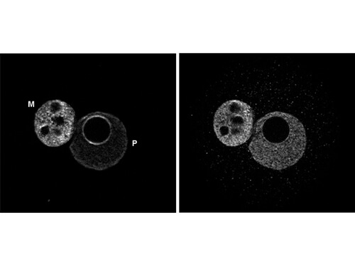
Fluorescence in situ Hybridization (FISH) of Mouse Anti-5-mC antibody. Cells: mouse embryos cells. Treatment: none. Fixation: 4% PFA in PBS at 4C overnight. Block: 0.5% Triton X-100 for 30 min, PBS containing 2% BSA, then 2N HCl. Primary antibody: (Left) anti-5-mC antibody, (Right) anti-single stranded DNA antibody, at 1:200 for 1 hr at RT. Secondary antibody: anti-Mouse CY5 secondary antibody at 1:10,000 for 45 min at RT. Staining: (left) both the maternal (M) and paternal (P) pronuclei are stained with 5-mC antibody. (right) staining of the DNA with an anti-single stranded DNA antibody.
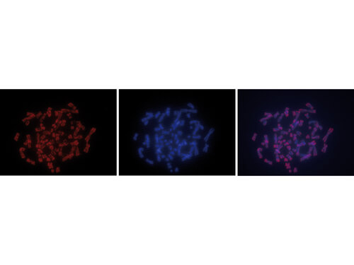
Fluorescence in situ Hybridization (FISH) of Mouse Anti-5-mC antibody. Cells: metaphase chromosomes from HeLa cells. Treatment: colcemid (0.1 µg/ml) for 1 - 2 hours. Fixation: ethanol/glacial acetic acid -20C overnight then treated with 2N HCl for 30min at RT. Block: 0.1% Triton X-and 1%BSA. Primary antibody: 5-mC antibody at 1:1000 for 1 hr at RT. Secondary antibody: goat anti-Mouse Alexa594 secondary antibody at 1:10,000 for 45 min at RT. Staining: 5-mC antibody as red signal (left), with blue DAPI (middle), and images merged (right).
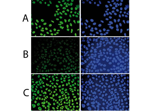
Immunofluorescence Microscopy results of Mouse anti-5-mC antibody. Tissue: human osteosarcoma cells (U2OS). Fixation: 2.5% PFA for 30 min. Antigen retrieval: 0.5% Triton X-100 for 1 hour and treated with 2N HCl for 1 hour followed by 2 x 5 minutes with 0.1 M borate buffer to depurinate the DNA. Primary antibody: 5-mC antibody at 1:500. Secondary antibody: Goat anti-Mouse Alexa488 secondary antibody. Staining: (A): 5-mC antibody as green fluorescent signal (left) with DAPI (right). (B): 5-cM antibody with 50 µM mCTP. (C): 5-cM antibody with 50 µM hmCTP.
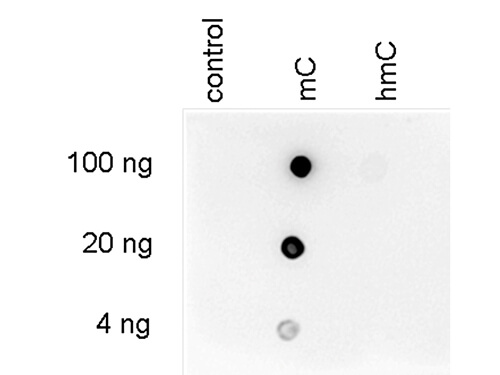
Dot Blot results of Mouse anti-5-mC monoclonal antibody. Antigens: C control, mC, and hmC forms of the immunizing peptide. Load: 100 ng, 20 ng, and 4 ng as indicated. Primary antibody: 5-mC antibody at 1:600 for 45 min at 4C. Secondary antibody: goat anti-mouse Dylight(TM)488 secondary antibody at 1:10,000 for 45 min at RT. Block: 5% BLOTTO overnight at 4C.
* Mehrwertsteuer und Versandkosten nicht enthalten. Irrtümer und Preisänderungen vorbehalten