0.02 M Potassium Phosphate, 0.15 M Sodium Chloride, pH 7.2
Formulierung:
Lyophilized
Target-Kategorie:
Human
Antibody Type:
Primary Antibody
Application Verdünnung:
ELISA: 1:20,000 - 1:100,000, IHC: 1:1,000 - 1:5,000, IF Microscopy: User Optimized, WB: 1:2,000 - 1:10,000
Anwendungsbeschreibung:
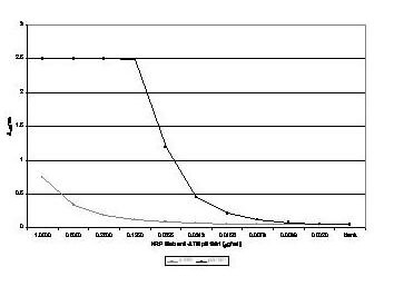
This antibody was tested by ELISA and western blotting against both the native and recombinant forms of the protein. This reagent may also be suitable other biotin-streptavidin based assays.
Anti ATM Antibody showing overlay of anti-ATM pS1981 staining.
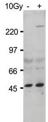
Western Blot of Rocklands Protein A Purified Mab anti-ATM Protein Kinase pS1981. Lane 1: HEK293 cells treated with doxorubicin pre-incubation of peptide with 50 µg of immunizing phospho peptide negates specific staining. Lane 2: HEK293 cells treated with doxorubicin. A 370 kDa band corresponding to phosphorylated ATM is detected (lane 2). The lysate was prepared with HALT phosphatase inhibitor (Pierce). Load: ~30µg. Primary antibody: anti-ATM Protein Kinase pS1981 diluted 1:500 overnight at 4C. Secondary Antibody: IRDye(TM)800 conjugated Gt-a-Mouse IgG [H&L] (code 610-132-121) at 1:10,000 for 40 min at room temperature. LICORs Odyssey Infrared Imaging System was used to scan and process the image. Other detection systems will yield similar results.
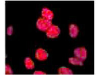
Anti ATM Antibody showing overlay of anti-ATM pS1981 staining. Cells were fixed 15 min after 5 Gy (IR+) of irradiation, then labeled with antibody. See Kitagawa et al. for additional details.
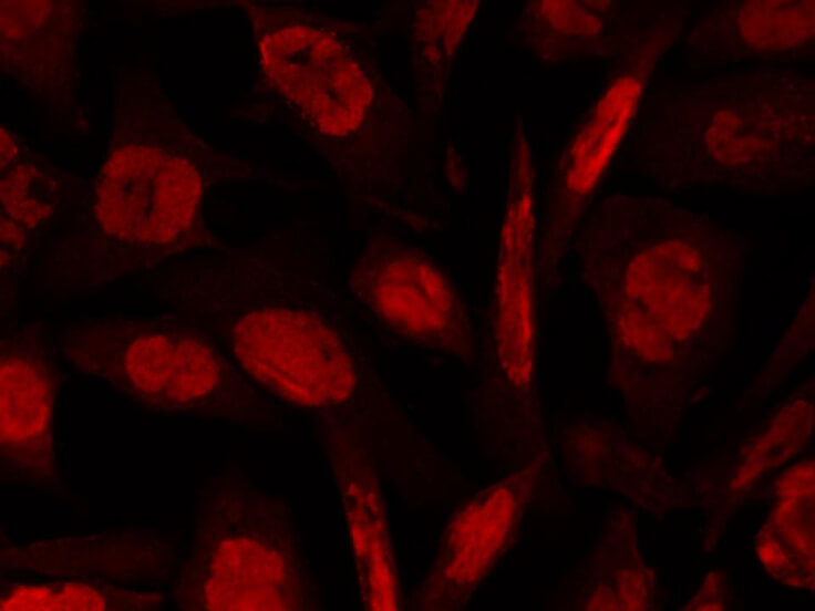
Rocklands anti-ATM pS1981 mouse monoclonal antibody (Catalog 200-301-400) detects ATM phosphorylated on Ser 1981 by Indirect immunofluorescence microscopy. Shown are hTCEpi cells (courtesy of Dr. Danielle Robertson) infected with HSV-1 at MOI 5.0 and fixed at 8 hpi with 3% paraformaldehyde/2% sucrose for 10 min. After rinsing, cells were permeabilized with 0.5% Triton X-100 for 5 min, blocked with 3% BSA for 30 min, and stained with Rocklands primary anti-ATM pS1981 antibody overnight at 5 µg/mL (1:200). Secondary staining was performed with Alexa Fluor 594 anti-mouse antibody. Images were taken with Olympus AX70 compound epifluorescence microscope equipped with Spot RT Slider camera. Experiment was performed by Oleg Alekseev in the laboratory of Dr. Jane Azizkhan-Clifford at Drexel University College of Medicine.
Anti ATM Antibody showing overlay of anti-ATM pS1981 staining. Cells were fixed 15 min after 5 Gy (IR+) of irradiation, then labeled with antibody. See Kitagawa et al. for additional details.
* Mehrwertsteuer und Versandkosten nicht enthalten. Irrtümer und Preisänderungen vorbehalten