Goat Anti Human IgG DyLight 549(TM) Conjugated Antibody, Goat Anti-Human IgG Antibody DyLight 549(TM) conjugation
Klonalität:
Polyclonal
Konzentration:
1.0 mg/mL by UV absorbance at 280 nm
Puffer:
0.02 M Potassium Phosphate, 0.15 M Sodium Chloride, pH 7.2
Formulierung:
Lyophilized
Target-Kategorie:
Human
Antibody Type:
Secondary Antibody
Application Verdünnung:
FLISA: >1:20,000, IF Microscopy: >1:5,000, WB: >1:10,000
Anwendungsbeschreibung:
Anti-Human IgG (H&L) DyLight 549 has been tested by ELISA and dot blot and is designed for immunofluorescence microscopy, fluorescence based plate assays (FLISA) and fluorescent western blotting. This product is also suitable for multiplex analysis, incl
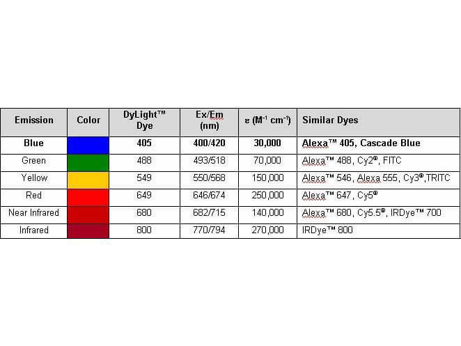
Properties of DyLight(TM) Conjugates.
Western Blot analysis of IgG antibodies againstR.helveticawhole cell antigen. Lane A-P demonstrates the lipopolysaccaride ladders and specific reactions againstR.helveticaproteins in the 110-150-kDa region for serum 2 for patients (Lane) V16(A), V43(B), V46 (C)(Area V), S71(D), S72(E), S75(F), V6(G) (Area Ö), K7(H), K9(I), K14(J), K46(K) K56(L) (Area K), A13(M), A16(N), A23(O), A35(P) (Area A) in titres 1:200. Lane P(h) demonstrates specific proteins and the lipopolysaccharide (LPS) ladders reacting with a human antiserum from a patient diagnosed with rickettsial infection and N(h) a healthy negative blood donor. Mw = molecular weight marker. ''Fig 2 is compiled of four figure panels representing the groups of lanes that originated from different gels/blots (Gel A-D). The short vertical lines of ''Fig 2 divide the individual non-adjacent lanes in the gels. The original analyses are presented inS1-S4Figs with Gels A-D as Supporting Information. Fig 2. PMID: 27846275.
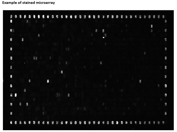
702 Peptides are printed in duplicates randomly distributed on the microarray. Control peptides (HA and FLAG controls) are located in a square surrounding the peptides of interest. As secondary antibody DyLight(TM) 549 conjugated goat anti-human IgG antibody and for the FLAG control peptide a mouse anti-FLAG-Cy3 antibody were used, microarrays were read using a Fujifilm Life Science FLA-5100 imaging system with a SHG 532nm (green) diode laser and an LPG filter. Fig e1. PMID: 26894206.
Western Blot analysis of IgG and IgM antibodies againstR. helveticawhole cell antigen demonstrates the lipopolysaccaride (LPS) ladders and specific reactions againstR. helveticaproteins in the 110-150-kDa region in serum for IgG for patients 1, 32 and 33 and for IgM for patients 3, 8, 10, 17, 20 and 22 in dilution 1:200. Lane P(h) demonstrates specific proteins and the LPS ladders reacting with a positive human serum and P(r) with a polyclonal rabbit antiserum. N(h) represent a negative human serum control. Secondary antibody Anti-human IgG DyLight(TM)549 (p/n 609-142-123 ) and Anti-human IgM DyLight(TM) 549 (p/n 609-142-007). Figure 1. PMID: 34712390.
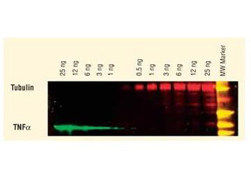
DyLight(TM) dyes can be used for two-color western blot detection with low background and high signal. Anti-tubulin was detected using a DyLight(TM) 549 conjugate. Anti-TNFa was detected using a DyLight(TM) 649 conjugate. The image was captured using the Typhoon(TM) 9410 Imaging System.
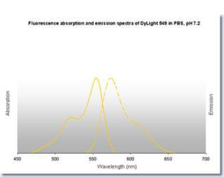
DyLight(TM) 549 Fluorescence Spectra.
* Mehrwertsteuer und Versandkosten nicht enthalten. Irrtümer und Preisänderungen vorbehalten