Goat Anti Human IgG DyLight 680(TM) Conjugated Antibody, Goat Anti-Human IgG Antibody DyLight 680(TM) conjugation
Klonalität:
Polyclonal
Konzentration:
1.0 mg/mL by UV absorbance at 280 nm
Puffer:
0.02 M Potassium Phosphate, 0.15 M Sodium Chloride, pH 7.2
Formulierung:
Lyophilized
Target-Kategorie:
Human
Antibody Type:
Secondary Antibody
Application Verdünnung:
FLISA: >1:20,000, IF Microscopy: >1:5,000, WB: >1:10,000
Anwendungsbeschreibung:
Anti-Human IgG (H&L) DyLight 680 has been tested by dot blot and is designed for immunofluorescence microscopy, fluorescence based plate assays (FLISA) and fluorescent western blotting. This product is also suitable for multiplex analysis, including mult
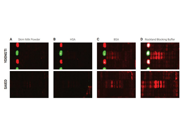
Selected sections of the PEPperCHIP Peptide Microarrays after assay with different blocking reagents. The microarrays were blocked for 30 minutes with either 2% skim milk powder (A), 1% HSA (B), 1% BSA (C) or 100% Rockland Blocking Buffer [p/n MB-070] (D), respectively. A human serum sample was assayed at dilution 1:200, followed by detection with secondary goat Anti-Human IgG (H+L) DyLight(TM) 680 Antibody [p/n 609-144-123]. Red spots = sample responses and polio control peptides, green spots = HA control peptides. The underlying binding motifs of the respective sections are indicated on the left.
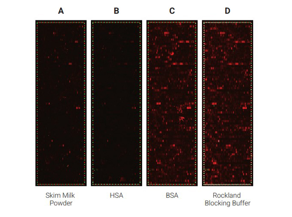
Comparison of the performance of different blocking reagents in epitope mappings with PEPperCHIP Peptide Microarrays.The PEPperCHIP Peptide Microarrays were blocked for 30 minutes with either 2% skim milk powder (A), 1% HSA (B), 1% BSA (C) or 100% Rockland Blocking Buffer [p/n MB-070] (D). A human serum sample was assayed at dilution 1:200, followed by detection with secondary goat anti-Human IgG (H+L) DyLight(TM) 680 Antibody [p/n 609-144-123] and a control anti-HA (12CA5)-DyLight(TM) 800 Antibody. Red spots = sample IgG response and frame of polio control peptides, green spots = frame of HA control peptides.
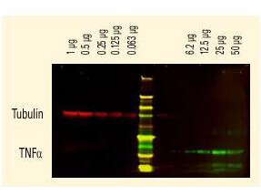
DyLight(TM) dyes can be used for two-color Western Blot detection with low background and high signal. Anti-tubulin was detected using a DyLight(TM) 680 conjugate. Anti-TNFa was detected using a DyLight(TM) 800 conjugate. The image was captured using the Odyssey Infrared Imaging System developed by LI-COR.
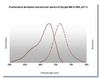
DyLight(TM) 680 Fluorescence Spectra.
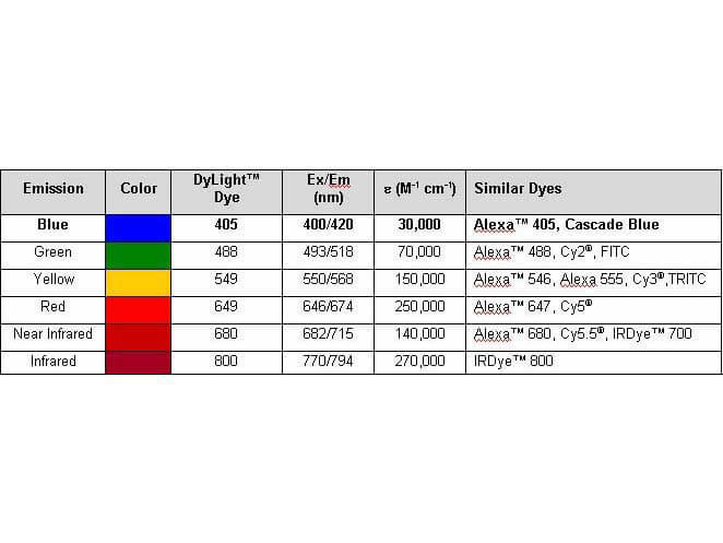
Properties of DyLight(TM) Conjugates.
* Mehrwertsteuer und Versandkosten nicht enthalten. Irrtümer und Preisänderungen vorbehalten