0.02 M Potassium Phosphate, 0.15 M Sodium Chloride, pH 7.2
Formulierung:
Lyophilized
Target-Kategorie:
Mouse
Antibody Type:
Secondary Antibody
Application Verdünnung:
FLISA: >1:20,000, IF Microscopy: >1:5,000, WB: >1:10,000
Anwendungsbeschreibung:
This product is designed for immunofluorescence microscopy, fluorescence based plate assays (FLISA) and fluorescent western blotting. This product is also suitable for multiplex analysis, including multicolor imaging, utilizing various commercial platfor
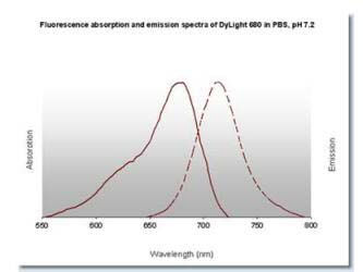
DyLight(TM) 680 Fluorescence Spectra.
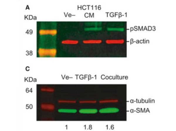
TGF-beta-mediated crosstalk between pericytes and CRC cells modulates pericyte secretome. (A) Incubation in HCT116 CM for 1h induces SMAD3 phosphorylation in PC, as assessed by western blot. Exogenous recombinant TGF-beta (10ngmL-1) was used as a positive control, and beta-actin was used as loading control (n=3). Fig. 5. PMID: 32767843.
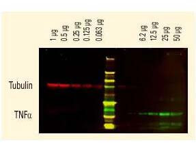
DyLight(TM) dyes can be used for two-color western blot detection with low background and high signal. Anti-tubulin was detected using a DyLight(TM) 680 conjugate. Anti-TNFa was detected using a DyLight(TM) 800 conjugate. The image was captured using the Odyssey Infrared Imaging System developed by LI-COR.
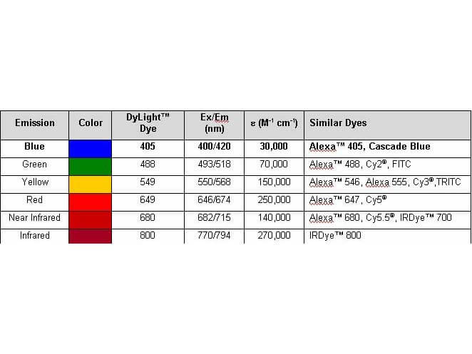
Properties of DyLight(TM) Fluorescent Dyes.
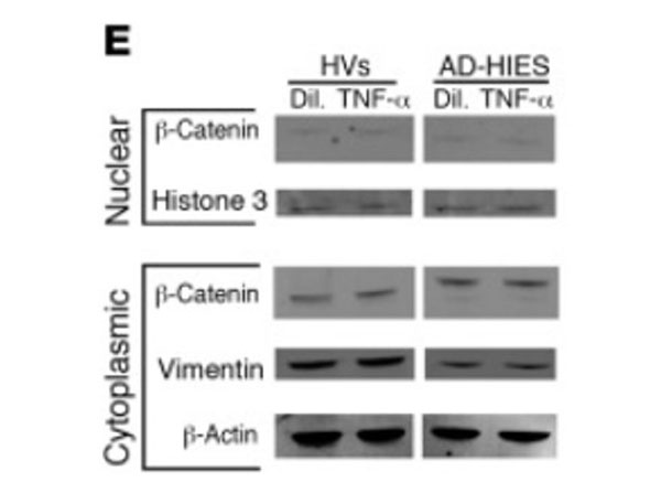
Skin cell cultures from patients with AD-HIES display TNF-alpha-sensitive defects in wound healing.(E) Western blot of nuclear and cytoplasmic fractions from KCs obtained from HVs and AD-HIES patients (KCs were cultured with TNF-alpha or diluent). Figure 3. PMID: 30035749.
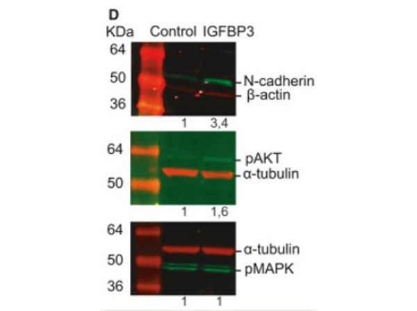
Insulin-like growth factor-binding protein 3 increases CRC cell migration and invasion through Akt activation. (D) Treatment with 50ngmL-1IGFBP-3 for 72h promotes the expression of N-cadherin in HCT116 cells as assessed by western blot (top panel). Phosphorylation status of Akt (middle panel) and MAPK (bottom panel) in HCT116 cells treated with 50ngmL-1IGFBP-3 for 15min. Representative images of three independent experiments (n=3). Numbers indicate the expression fold change relative to the loading control. Fig. 7. PMID: 32767843.
* Mehrwertsteuer und Versandkosten nicht enthalten. Irrtümer und Preisänderungen vorbehalten