Rabbit IgG Fc Antibody DyLight(TM) 680 Conjugated, DL680, Goat, Polyclonal
Artikelnummer:
ROC-611-144-003
Hersteller Artikelnummer:
611-144-003
Alternativnummer:
ROC-611-144-003
Hersteller:
Rockland Immunochemicals
Wirt:
Goat
Kategorie:
Antikörper
Spezies Reaktivität:
Rabbit
Immunogen:
Rabbit IgG F(c) fragment
Konjugation:
DL680
Alternative Synonym:
Goat Anti Rabbit IgG F(c) DyLight 680(TM) Conjugated Antibody, Goat Anti-Rabbit IgG Fc Fragment Antibody DyLight 680(TM) conjugation, Goat Anti Rabbit IgG Fc Antibody DyLight 680(TM) conjugated
Klonalität:
Polyclonal
Konzentration:
1.0 mg/mL by UV absorbance at 280 nm
Puffer:
0.02 M Potassium Phosphate, 0.15 M Sodium Chloride, pH 7.2
Formulierung:
Lyophilized
Target-Kategorie:
Rabbit
Antibody Type:
Secondary Antibody
Application Verdünnung:
FLISA: >1:20,000, IF Microscopy: >1:5,000, WB: >1:10,000
Anwendungsbeschreibung:
The emission spectra for this DyLight(TM) conjugate match the principle output wavelengths of most common fluorescence instrumentation. This product is designed for immunofluorescence microscopy, fluorescence based plate assays (FLISA) and fluorescent weste
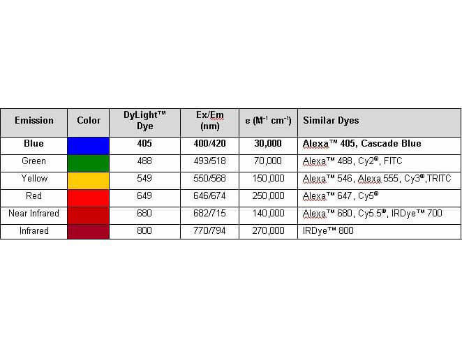
Properties of DyLight(TM) Conjugates.
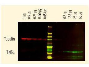
DyLight(TM) dyes can be used for two-color Western Blot detection with low background and high signal. Anti-tubulin was detected using a DyLight(TM) 680 conjugate. Anti-TNFa was detected using a DyLight(TM) 800 conjugate. The image was captured using the Odyssey Infrared Imaging System developed by LI-COR.
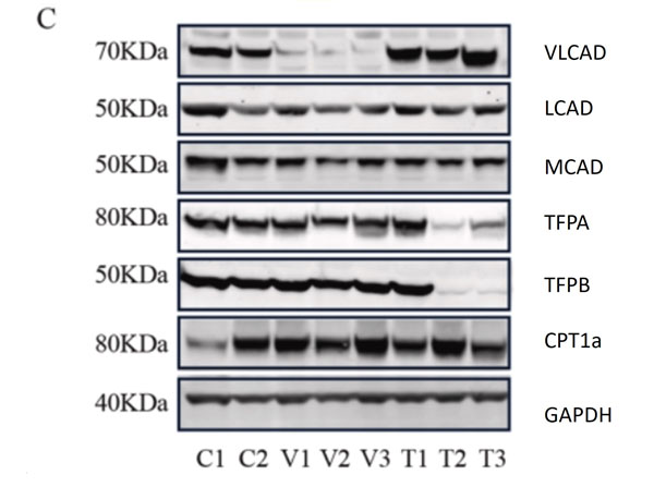
C.Representative western blots, original blots are shown in(supplementary Fig S8-9). And densitometric quantification of relative protein levels from western blots. Data are depicted as meanSD, n=3, **P<0.01, ***P<0.001 and ****P<0.0001 by one-way ANOVA. Intracellular transport, activation, mitochondrial transport, beta-oxidation, carnitine shuttle, and auxiliary proteins. The primary antibodies used as follows: VLCAD 1:1000, MCAD 1:1000, LCAD 1:1000, TFPa 1:500, TFPb 1:3000, CPT1alpha 1:1000, and GAPDH 1:30,000 dilutions overnight at 4C. The membranes were then incubated with fluorescent conjugated secondary antibodies for 1h, DyLight 800 conjugated goat Anti-Rabbit IgG (611-145-002), DyLight 680 conjugated goat Anti-Rabbit IgG (611-144-003), DyLight 800 conjugated goat Anti-Mouse IgG (610-145-002), and DyLight 680 conjugated donkey Anti-Mouse IgG (610-744-124). Fig 1. PMID: 33725513.
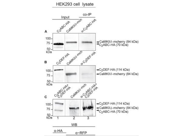
Immunoprecipitation and western blot show interaction of otoferlin with CaMKIIdelta.(A-C)Two HA-tagged mouse otoferlin fragments, C2ABC (aa 1-632 inNP_001093865, 70 kDa) and C2DEF (aa 933-1920, 114 kDa) were co-transfected with mcherry-tagged mouse CaMKIIdelta into HEK293 cells. Transfections were performed either with otoferlin C2ABC and CaMKIIdelta (A, Input Lane 1 and 2), otoferlin C2DEF and CaMKIIdelta (B, Input Lane 1 and 2) or in the presence of both C2ABC and C2DEF fragments and CaMKIIdelta (C, Input Lane 1 and 2). Co-immunoprecipitations of C2ABC-HA and C2DEF-HA were conducted from HEK293 cell lysates using anti-HA antibodies (p/n 600-401-384). CaMKIIdelta-mcherry was detected in the eluate using an anti-RFP (red fluorescent protein) antibody (p/n 200-301-379) (A-C, Lane 3), indicating that CaMKIIdelta co-precipitated with recombinant otoferlin fragments. Secondary anti-rabbit Dylight680 (p/n 611-144-003) and anti-mouse Dylight800 antibodies (610-145-003) (1:10,000). FIGURE 5. PMID: 29046633.
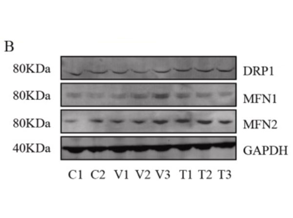
Assessment of mitochondrial fusion and fission.B.Representative western blots (original blots are shown insupplementary Fig. S10) and quantification of MFN1/2 and DRP1. No significant changes in the relative levels of proteins that facilitate mitochondrial fusion (MFN1/2) and fission (DRP1) between non-disease (control) and mutant primary fibroblasts. Data are depicted as meanSD, n=3. The primary antibodies used as follows: MFN1 1:400, MFN2 ( 1:400, DRP1 1:100 and GAPDH 1:30,000 dilutions overnight at 4C. The membranes were then incubated with fluorescent conjugated secondary antibodies for 1h, DyLight 800 conjugated goat Anti-Rabbit IgG (611-145-002), Antibody DyLight 680 conjugated Anti-Rabbit IgG made in goat (611-144-003), DyLight 800 conjugated goat Anti-Mouse IgG (610-145-002), and DyLight 680 conjugated donkey Anti-Mouse IgG (610-744-124). Fig 3. PMID: 33725513.
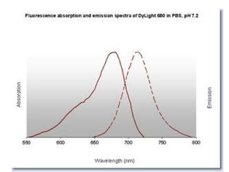
DyLight(TM) 680 Fluorescence Spectra.
* Mehrwertsteuer und Versandkosten nicht enthalten. Irrtümer und Preisänderungen vorbehalten