ELISA: User Optimized, FLISA: User Optimized, IHC: User Optimized, IF Microscopy: User Optimized, IP: User Optimized, WB: User Optimized
Anwendungsbeschreibung:
BOVINE SERUM ALBUMIN 30% Solution is suitable for use in protease sensitive assays such as RIA, EIA and nucleic acid hybridization, use as a stabilizing agent for proteins and enzymes, including dilute solutions of antibody, and use as a blocking agent t
Bovine Serum Albumin 30% Solution
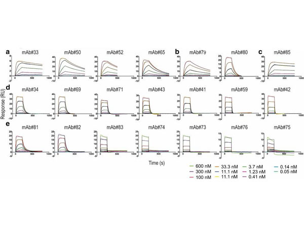
Surface plasmon resonance sensorgrams representing the binding of basigin extracellular domain to captured anti-basigin monoclonal antibodies. The fitted curves used for equilibrium dissociation constant (KD) calculations were obtained using the Langmuir 1:1 binding model in the Biacore S200 evaluation software and plotted in GraphPad Prism (colored in black). (a) bin A, (b) bin D, (c) bin AD, (d) bin B, and (e) bin C.At the end of each cycle, the sensor surface was regenerated in 3M MgCl2for 30s. 1*HBS-P+with 1mg/ml bovine serum albumin (BSA, p/n BSA-30) was used as running buffer for the kinetic analyses. mAbmonoclonal antibody,RUresponse unit. Figure 3. PMID: 32884039.
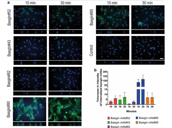
Internalization of basigin monoclonal antibodies (mAbs) in hCMEC/D3 cells. (a) Representative confocal images of selected basigin mAbs exposed to hCMEC/D3 cells for 10 and 30min. Basigin mAbs in green and nuclei visualized by Hoechst in blue. The pictures are the maximum projection of the z-stack, and the XZ projection is below the pictures. Scale bar 30µm. (b) Quantification of intracellular spots using Cellomics Arrayscan after acid stripping and staining. The intensities are normalized to the negative control and plotted as the percentage increase in spot intensity per cell withstandard error of the mean (SEM). Secondary antibody diluted in PBS with 2% BSA (p/n BSA-30).Figure 6. PMID: 32884039.
* Mehrwertsteuer und Versandkosten nicht enthalten. Irrtümer und Preisänderungen vorbehalten