normal goat serum, NGS, blocking serum, goat serum for bioassay, control goat serum, non-sterile serum from goat, non-sterile goat serum
Konzentration:
104 mg/mL by Refractometry
Puffer:
None
Quelle:
Goat
Formulierung:
Liquid
Application Verdünnung:
ELISA: User Optimized, FLISA: User Optimized, Flow Cytometry: User Optimized, IHC: User Optimized, IF Microscopy: User Optimized, IP: User Optimized, WB: User Optimized
Anwendungsbeschreibung:
pH: normal Immunoelectrophoresis: normal Hemoglobin: normal IgG Concentration: normal
Normal Goat Serum can be used as a blocking agent to treat plastic surfaces, membrane or tissue after they have been sensitized with primary antibody or antigen. It provides an alternative to bovine serum albumin (BSA) and non-fat dry milk. It is effective in reducing nonspecific binding of proteins to reaction surfaces, thereby maximizing signal-to-noise ratio. This blocking agent is recommended for use in immunoassays where the primary antibody was produced in goat, as a source of non-specific serum protein or on tissue for immunohistochemical applications.
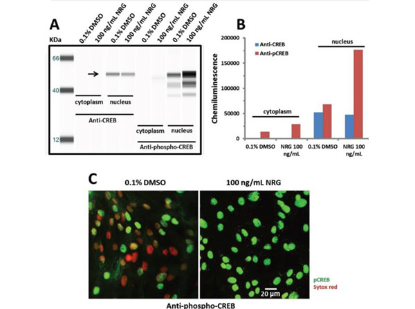
Nuclear specific expression of CREB and activation after neuregulin-1beta (NRG) treatment as shown by Wes(TM) Simple Western (A) and IN Cell Analyzer 2000 microscopic cell imaging (B). Cells were cultured more than 14 days prior to testing, and equilibrated to serum-free media for about 45 minutes before being exposed to 100 ng/mL NRG for 30 minutes. Upon completion of NRG treatment, cells were lysed and the fraction of cytoplasmic or nuclear proteins was extracted using the NE-PER Nuclear and Cytoplasmic Extraction kit for Wes(TM) Simple Western analysis (A). A band at 53 KDa was detected by both the pan- and phospho-CREB antibodies in nuclear, but not in cytoplasmic extracts (arrow). (B) Total CREB in the nucleus was not altered by treatment with 100 ng/mL NRG, whereas NRG treatment increased phosphorylated CREB by approximately 160% compared to the 0.1% DMSO group. Cells were fixed and stained with anti-phospho-CREB and Sytox Red (nuclear stain) for immunofluorescence using the IN Cell Analyzer 2000 system for imaging (C). Cells were treated with 0.1% DMSO or 100 ng/mL NRG as described for panel A. Images were acquired using the IN Cell Analyzer 2000 instrument and image analysis was performed using the IN Cell Workstation. Nuclear staining for phospho-CREB was clearly increased after treatment with 100 ng/mL NRG as seen by the increase in pCREB imaged in the green channel. Note the lack of phospho-CREB in many nuclei that stain only in the red (Sytox Red) channel in the DMSO control image. Based on analyzing more than 3,400 cells per group, treatment with NRG increased the average nuclear intensity of phospho-CREB by approximately 94% compared to the 0.1% DMSO treated cells, similar to the Wes data. 3% goat serum (p/n D204-00-0050) incubated for 1 hour at room temperature to block non-specific binding. Figure 16. PMID: 26331525.
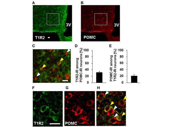
Distribution of T1R2 on POMC neurons in the ARC. Blocked with 1.5% normal goat serum (p/n D204-00-0050). T1R2 (A)and POMC(B)immunofluorescent images and their merged image with high magnification(C).(D)Percentage of T1R2-imunoreactive (IR) neurons among POMC-IR neurons in the ARC (n= 3 brains).(E)Percentage of POMC-IR neurons among T1R2-IR neurons in the ARC (n= 3 brains). Confocal images of T1R2(F)and POMC(G)immunofluorescent and their merged image(H). White arrowhead indicates non-colocalizing POMC-IR neuron, and yellow arrowhead indicates a colocalizing neuron. Scale bar, 20 µm. Graphs show mean SEM. Figure 6. PMID: 27877104.
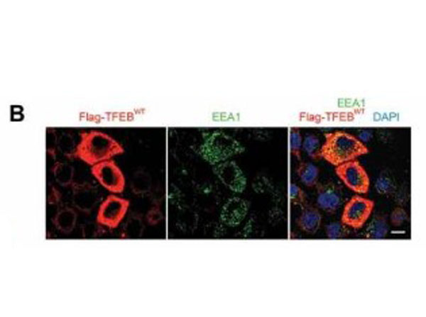
Overexpression of TFEB stimulates cellular endocytosis. (b) Immunofluorescence images represent HeLa cells transiently transfected with WT Flag-TFEB for 48h and immunostained for EEA1. Flag-TFEB-expressing cells showed a significant increase in the levels of EEA1-positive endosomes as compared to untransfected, neighboring cells. HEK293T cells transiently transfected with constitutively active TFEBS211Aalso showed an increased level of EEA1 expression. Cultures were rinsed in PBS and blocked in 5% normal goat serum (p/n D204-00-0100), 0.5% BSA, and 0.5% Triton X-100 in PBS for 45min at room temperature. Fig 1. PMID: 30145926.
* Mehrwertsteuer und Versandkosten nicht enthalten. Irrtümer und Preisänderungen vorbehalten