
Cyclin Antibody Sampler Kit
Artikelnummer:
ROC-K911
- Bilder (5)

| Artikelname: | Cyclin Antibody Sampler Kit |
| Artikelnummer: | ROC-K911 |
| Hersteller Artikelnummer: | K911 |
| Alternativnummer: | ROC-K911 |
| Hersteller: | Rockland Immunochemicals |
| Kategorie: | Kits/Assays |
| Applikation: | IHC, WB |
| Konjugation: | Unconjugated |
| Alternative Synonym: | Anti-Cyclin A Antibody, Anti-Cyclin B1 Antibody, Anti-Cyclin D1 Antibody, Anti-Cyclin E Antibody, Goat Anti-Rabbit IgG Antibody, antibody sample kit |
| Formulierung: | Liquid (sterile filtered) |
| Application Verdünnung: | ELISA: 1:2,000 - 1:10,000, IP: 1:100, WB: 1:500 - 1:5,000 |
| Anwendungsbeschreibung: | Each kit contains one unit of the following products: (100 µL) Anti-Cyclin A (RABBIT) Antibody (100 µL) Anti-Cyclin B1 (RABBIT) Antibody (100 µL) Anti-Cyclin D1 (RABBIT) Antibody (100 µL) Anti-Cyclin E (RABBIT) Antibody (100 µg) Anti-RABBIT IgG (H&L) (GO |
|
|
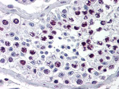
Rocklands Anti-Cyclin A antibody was diluted 1:500 to detect Cyclin A in human testes tissue. Tissue was formalin fixed and paraffin embedded. No pre-treatment of sample was required. The image shows the localization of antibody as the precipitated red signal with a hematoxylin purple nuclear counter stain. |
|
|
|
|
|
This product is assembled as a kit. See attached protocol or CofA for further details. |
|
|
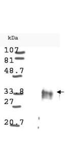
Western blot analysis is shown using Rocklands anti-Cyclin D1 antibody to detect Human Cyclin D1 present in asynchronous HN30 cell lysates. HN30 cells, are from head and neck cancer cells that over express cyclin B1 and D1. Comparison to a molecular weight marker indicates a band of ~34 kDa corresponding to the expected molecular weight for the protein (arrowhead). The blot was incubated with a 1:500 dilution of the antibody at room temperature. Detection occurred using a 1:10,000 of HRP conjugated Goat-a-Rabbit IgG 611-103-122 and chemiluminescence reagent with a 1-min exposure time. Other detection systems will yield similar results. Personal communication Luca Cote. |
|
|
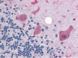
Rocklands anti-Cyclin B1 antibody was diluted 1:500 to detect Cyclin B1 in human brain cerebellum tissue. Tissue was formalin fixed and paraffin embedded. No pre-treatment of sample was required. The image shows the localization of antibody as the precipitated red signal, with a hematoxylin purple nuclear counter stain. |
Produktgarantie und fachkundiger Support
