Chicken Washed Pooled Cells, Chicken WPCs, Chicken Red Blood Cells, Chicken RBCs, erythrocytes
Puffer:
0.02 M Potassium Phosphate, 0.15 M Sodium Chloride, pH 7.2
Quelle:
Chicken
Formulierung:
Liquid
Anwendungsbeschreibung:
Chicken whole blood cells are used for complement titration, adsorption procedures, HA assays and for the preparation of stroma as particulate reagents.
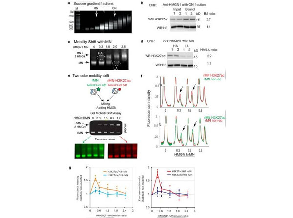
Preferential binding of HMGN to chromatin particles containing H3K27ac. aAgarose gel showing sucrose gradient fractionated salt stripped MEF chromatin particles. MN: mononucleosomes, ON: Oligonucleosome (mostly tri-penta nucleosomes).bWestern analysis of total ON (Input) and HMGN1 immunoprecipitated ON (bound) (c) Gel mobility shift -assay. Purified HMGN1 was added to salt stripped chicken erythrocyte MNs at the ratio indicated on top of each column. The MNs shifted at low HMGN1:MN were designated as high affinity (HA) while the MNs not shifted at high HMGN:MN were designated as low affinity (LA).dWestern analysis of HA and LA mononucleosomes (MN).eTwo color gel mobility shift assays of recombinant mononucleosomes (rMN). A mix of equal amounts of fluorescently Alexa 488 labeled rMN (green) and Alexa 647 labeled rMNH3K27ac (red) were incubated with various amounts of HMGN1, the mixture fractionated on native polyacrylamide gels, and the gels scanned to visualize and quantify either the red or green fluorescence. Shown is the experimental design and gel images visualized with red or green channels (f) Scan of the gels shown ineand of a similar gel in which the fluorescent labels are reve
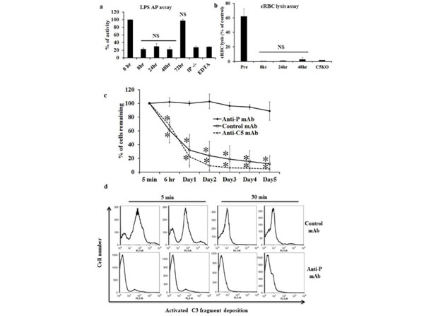
Therapeutic efficacy of anti-human P mAb 19.1 in a murine model of extravascular hemolysisa:Pharmacodynamics of mAb 19.1 in hP transgenic mice. Each mouse was treated with 0.5 mg mAb 19.1 (n=3 mice). Serum samples were collected before and at various time points after mAb treatment and assessed for LPS-dependent AP complement activity. At this dosage of mAb 19.1, AP complement activity was suppressed to background (P-/-) level for 2 days. EDTA: time 0 sample with EDTA (20 mM) added. NS, non-significant comparing 8, 24 and 48 hr samples with fP-/-or EDTA-treated serum, or comparing 72 hr sample with 0 hr sample. One-way ANOVA.b:Pharmacodynamics of anti-mouse C5 mAb (BB5.1) in hP transgenic mice. Each mouse was treated with 1mg of anti-mouse C5 mAb (n=3). Serum samples were collected before and at various time points after mAb treatment and assessed for lytic activity using antibody-sensitized chicken RBCs. C5 knockout (KO) mouse serum was used as a control for C5 inhibition. Percentage of chicken RBC (cRBC) lysis was normalized to a sample completely lysed by hypotonic shock in double distilled water. * p<0.0001, NS: non-significant compared with C5KO serum. One-way ANOVA.c.Effect of anti-hP mAb 19.1 on the survival of transfused CFSC-labeled DAF-/-/Crry-/-/C3-/-mouse erythrocytes in hP- transgenic mice. Recipient mice were treated with anti-hP mAb 19.1 (n=4) or an isotype control mAb (n=4) or anti-mouse C5 mAb (n=3) 6 hours prior to red blood cell transfusion and blood samples were taken at 5 min, 6 hrs and then daily for 5 days. The percentage of CFSC-labeled red blood cells was measured by FACS and normalized to that determined at 5 min (100%). Transfused DAF-/-/Crry-/-/C3-/-mouse red blood cells were rapidly eliminated in control mAb- or anti-C5 mAb-treated hP transgenic mice but such an outcome was prevented by mAb 19.1 treatment. * p< 0.001, Two-way ANOVA.d. FACS analysis of activated C3 fragment deposition on DAF-/-/Crry-/-/C3-/-RBCs 5 and 30 min after their transfusion into control mAb- or mAb 19.1-treated hP-Tg/P-/-mice (representative data from two recipient mice are shown). At both time points, C3 fragment deposition was significantly higher on RBCs transfused into control mAb-treated than mAb 19.1-treated hP-Tg/P-/-mice. The reason for the marked reduction in C3 fragment deposition on RBCs in control mAb-treated hP-Tg/P-/-mice between 5 and 30 min is unknown, but could be caused by C3 fragment degradation to C3d or shedding from the cell surface or rapid removal of the C3-opsonized cells. Data in a-c are presented as mean (SD) with indicated n numbers. Antibody-sensitized chicken RBC (p/n R401-0050) prepared by incubating the cells with a rabbit anti-chicken RBC antibody (p/n 103-4139). Fig 4. PMID: 29898960.
* Mehrwertsteuer und Versandkosten nicht enthalten. Irrtümer und Preisänderungen vorbehalten