Turkey Washed Pooled Cells, Turkey WPCs, Turkey Red Blood Cells, Turkey RBCs, erythrocytes
Puffer:
0.02 M Potassium Phosphate, 0.15 M Sodium Chloride, pH 7.2
Quelle:
Turkey
Formulierung:
Liquid
Anwendungsbeschreibung:
Complement titration, adsorption procedures, HA assays and for the preparation of stroma as particulate reagents.
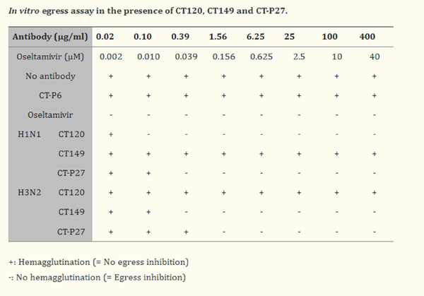
To address whether CT120, CT149, and CT-P27 could inhibit the egress step in the virus life cycle,in vitroegress assay was performed. MDCK cells were infected with A/Ohio/1983 (H1N1) or A/Philippines/2/1982 (H3N2). The infected cells were incubated to produce progeny viruses in the presence of CT120, CT149, or CT-P27. Released viruses were measured by HA assay since CT120, CT149, and CT-P27 could not inhibit hemagglutination even at 1.2 mg/mL concentration. The results show that CT120 and CT149 inhibited egress of H1N1 and H3N2 viruses, respectively, and CT-P27 inhibited egress of both H1N1 and H3N2 viruses. Viral antigens were mixed with 2-fold serially diluted antibodies in PBS, dispensed into 96-well plates, and incubated at 20-22C for 30 min. Next, 0.5% suspension of turkey erythrocytes (p/n R408-0050) was added to each well, and the mixture was incubated for 30 min at 20-25C before visual scoring for hemagglutination activity. Table 3. PMID: 32726321.
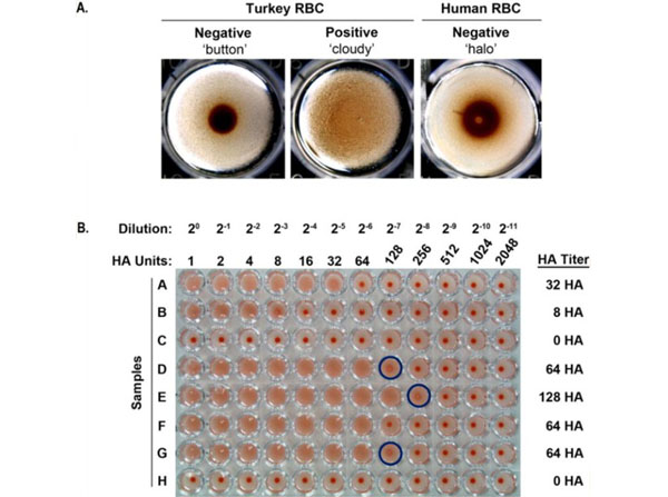
Hemagglutination (HA) assay (A) Samples lacking agglutinating activity (left panel) or containing IAV with agglutinating activity (center panel) were mixed with an equal volume of turkey RBCs (0.5%) in a U-bottomed microtiter plate and incubated for 30 minutes at room temperature, and a sample lacking agglutinating activity (right panel) was mixed with an equal volume of guinea pig [or human] RBCs (0.75%) in a U-bottomed microtiter plate and incubated for 1 hour at room temperature. At the end of each incubation period, 4X magnified images of individual microtiter plate wells were captured using a tissue culture microscope fitted with a digital camera. The left panel shows a characteristic negative 'button result, the central panel shows the evenly distributed, 'cloudy appearance of a positive agglutination result, and the right panel shows a thick ring of cells, i.e. a 'halo, negative result. (B) Samples containing IAV with agglutinating activity (see rows A, B, and D-G) or lacking IAV with agglutinating activity (see rows C and H) were subjected to an HA assay. Samples were 2-fold serially diluted (20- 211) in a 50-µl final volume, and then mixed with an equal volume of turkey RBCs (0.5%) in a U-bottomed microtiter plate. The entire microtiter plate was photographed after 30 minutes incubation at room temperature. The dilutions for each column and the corresponding HA units are indicated at the top of the panel, and the HA titer for each sample is indicated to the right. Wells exhibiting partial agglutination are indicated by dark blue circles. Turkey red blood cells (RBCs) (p/n R408-0050) and Guinea pig red blood cells (RBCs) (p/n R402-0050). Figure 4. PMID: 25321410.
* Mehrwertsteuer und Versandkosten nicht enthalten. Irrtümer und Preisänderungen vorbehalten