SA, S avidin, streptococcus avidin, streptavidin AT425, ATTO 425, ATTO-TEC 425, STREPTAVIDIN ATTO 425 Conjugated
Konzentration:
1.0 mg/mL by UV absorbance at 280 nm
Puffer:
0.02 M Potassium Phosphate, 0.15 M Sodium Chloride, pH 7.2
Formulierung:
Lyophilized
Application Verdünnung:
FLISA: >1:20,000, IF Microscopy: >1:5,000, WB: >1:10,000
Anwendungsbeschreibung:
Streptavidin ATTO 425 has been tested by dot blot, western blot, and immunofluorescence. The emission spectra for this ATTO conjugate matches the principle output wavelengths of most common fluorescence instrumentation.
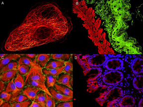
Atto Dye-Immunofluorescence microscopy ATTO dyes can be used for multicolor immunofluorescent detection with low background and high signal. Examples shown are: A. Tubulin in PtK2- male Rat Kangaroo Kidney Epithelial Cells was detected using ATTO 532 labeled secondary antibody. B. Muscle alpha-actin was stained with a mouse primary antibody and ATTO 488 anti-mouse IgG (green) while Cytokeratin was stained with polyclonal rabbit anti-cytokeratin and ATTO 647N anti-rabbit IgG (red). C. HUVEC (Human umbilical vein endothelial cells were stained with anti- Vimentin-ATTO 532 (green), anti-E-Cadherin-ATTO 655 (red) and DAPI (blue). D. Rat colon sections were stained with Anti-Aquaporin 3-ATTO 594 antibody. Hoechst 33342 (blue) is used as counterstain.
Streptavidin ATTO 425 Conjugated
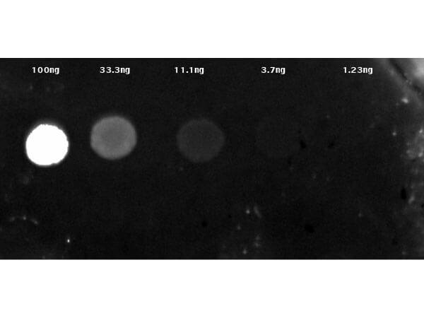
Dot Blot results of Streptavidin ATTO 425 Conjugate. Dots are Biotin: (1) 100ng, (2) 33.3ng, (3) 11.1ng, (4) 3.70ng, (5) 1.23ng. Primary Antibody: none. Secondary Antibody: Streptavidin ATTO 425 Conjugate at 1µg/mL in MB-070 for 1hr at RT. Imaged with BioRad ChemiDoc, CY2 filter.
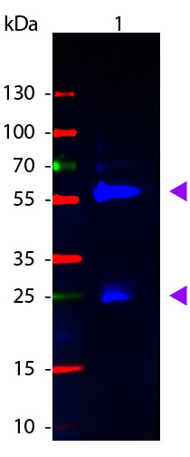
Western Blot of Atto 425 Conjugated Streptavidin. Lane 1: Biotin conjugated Guinea Pig IgG. Lane 2: None. Load: 50 ng per lane. Primary antibody: None. Secondary antibody: Atto 425 Conjugated Streptavidin at 1:1,1000 for 60 min at RT. Block: MB-070 for 30 min at RT. Predicted/Observed size: 28 & 55 kDa, 28 & 55 kDa for Guinea Pig IgG. Other band(s): None.
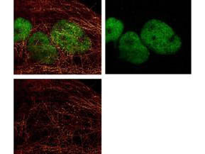
ATTO 425 conjugated anti-Mouse IgG was used to demonstrate 2 color STED immunofluorescence microscopy. Methanol fixed A431 cells were blocked with normal goat serum. The cells were then probed with 0.4 µg/mL final concentration of anti-a-tubulin and detected with 0.2 µg/mL ATTO 425 conjugated anti-MOUSE IgG [GOAT] (610-151-121) secondary antibody (colored RED). Also shown in this 2-color STED image is Rocklands Anti-HDAC-1 [RABBIT] (p/n 600-401-879) detected with DyLight(TM)488 conjugated Anti-RABBIT IgG [GOAT] secondary antibody (colored GREEN). Image courtesy of Myriam Gastard, Leica Microsystems, USA.
* Mehrwertsteuer und Versandkosten nicht enthalten. Irrtümer und Preisänderungen vorbehalten