Hela Cells, HeLa Lysate, Human Derived Whole Cell Lysate, Human Derived HeLa Whole Cell Lysate
Konzentration:
1.0 mg/mL by BCA assay
Puffer:
1X SDS-PAGE Sample Buffer (62.5 mM Tris HCl, 2% SDS, 10% Glycerol and 0.005% bromophenol blue, pH 6.8)
Quelle:
Human
Formulierung:
Liquid
Application Verdünnung:
WB: User Optimized
Anwendungsbeschreibung:
W09-000-364 has been tested by SDS-PAGE and western blot. Ready-to-use HeLa whole cell lysates are especially prepared as positive controls for separation by SDS-PAGE and subsequent western blot analysis. Lysates are prepared in denaturing buffer WITHOUT
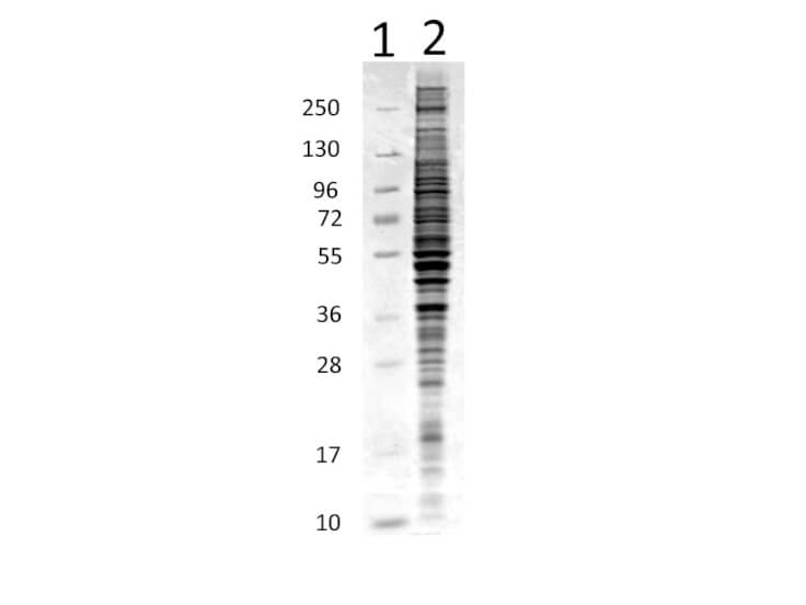
Coomassie stained SDS-PAGE of 10 µg of Human Derived HeLa Whole Cell Lysate separated using a 4-20% gradient gel under reducing conditions (lane 2). Molecular weight standards are shown in lane 1.
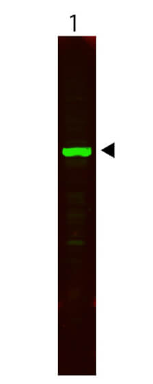
Western Blot showing detection of alpha tubulin in lane 1. HeLa Whole Cell Lysate (10 µg) was run on a 4-20% gel, then transferred to 0.45 µm nitrocellulose. After blocking with 1% BSA-TTBS (p/n MB-013, diluted to 1X) for 30 min at 20C, primary antibody was used at 1:2500 overnight at 4C. Anti-Rabbit IgG (H&L) (GOAT) antibody IRDye800CW (p/n 611-131-002) secondary antibody was used at 1:20,000 with Blocking Buffer for Fluorescent Western Blotting (p/n MB-070) and imaged on the LiCor Odyssey imaging system. Arrow indicates correct 50 kDa molecular weight position expected for alpha tubulin.
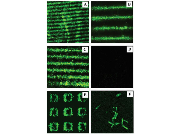
Fluorescence images acquired from TRE assay performed on ZnO NR arrays. The fluorescence images are (A) 1200 * 1200 m, (B) 400 * 400 m, (C) 120 * 120 m, (D) 120 * 120 m, (E) 60 * 60 m, and (F) 25*25 m in size. The underlying ZnO NR platforms used in assays A through D have striped patterns.(A-C) Confocal fluorescence data obtained from positive samples. The green fluorescence emission is clearly seen in these samples which signifies the detection of active telomerase and the successful incorporation of dNTPs in the extension of TS.(D) Typical fluorescence image collected from negative samples. No fluorescence emission is detected from these samples due to the lack of telomerase in the assay. The platforms used in assays E and F are open square and individual ZnO NRs, respectively.(E and F) Fluorescence images obtained from positive samples on (E) open square ZnO NR and (F) individual ZnO NR arrays. Negative assays were carried out by omitting either HeLa cell lysates (p/n W09-000-364) or dNTPs. Fig 3. PMID: 18468092.
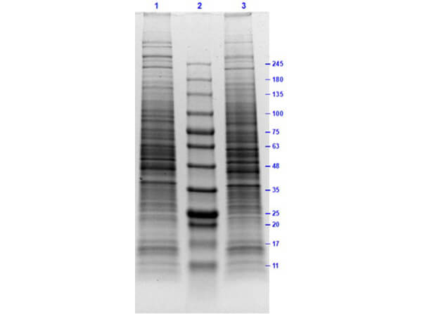
SDS PAGE Results of HeLa Whole Cell Lysates. Lane 1: HeLa Whole Cell Lysate Reduced [10µg]. Lane 2: Opal Prestained Molecular Weight Marker (p/n MB-210-0500). Lane 3: HeLa Whole Cell Lysate Non-Reduced [10µg]. 4-20% Gel, Coomassie Stained. Results show wide range of molecular weight bands with no signs of degradation.
* Mehrwertsteuer und Versandkosten nicht enthalten. Irrtümer und Preisänderungen vorbehalten