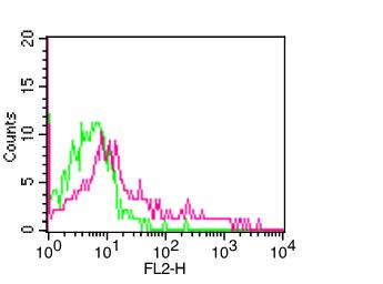
Fig-1: Cell surface flowcytometric staining of HLA-DP in hPBMC using 0.5 µg/ 10 6 cells of Anti-hHLA-DP (Clone:ABM54B6). Green represents isotype control, red represents anti-HLA-DP antibody. Goat anti-mouse PE conjugate was used as secondary antibody.
* VAT and and shipping costs not included. Errors and price changes excepted