
Connexin 43 Rabbit Polyclonal Antibody, Unconjugated
Catalog Number:
BYT-ORB10485
- Images (7)


| Article Name: | Connexin 43 Rabbit Polyclonal Antibody, Unconjugated |
| Biozol Catalog Number: | BYT-ORB10485 |
| Supplier Catalog Number: | orb10485 |
| Alternative Catalog Number: | BYT-ORB10485-50,BYT-ORB10485-100,BYT-ORB10485-200 |
| Manufacturer: | Biorbyt |
| Host: | Rabbit |
| Category: | Antikörper |
| Application: | FC, ICC, IF, IHC-Fr, IHC-P, WB |
| Species Reactivity: | Human, Mouse, Rat |
| Immunogen: | KLH conjugated synthetic peptide derived from human Connexin-43 (211-260/382 humanaa) |
| Conjugation: | Unconjugated |
| Alternative Names: | AVSD3, CMDR, CX43, EKVP, EKVP3, GJAL, HLHS1, HSS, ODDD, PPKCA, CXNK1, Cnx43, Cx43alpha1, Gja-1, Npm1, connexin43, CXA1_BOVIN, GJA1, Connexin-43 (Cx43), Vascular smooth muscle connexin-43, CXA1_CANLF, CXA1_HUMAN, Gap junction 43 kDa heart protein, CXA1_MOUSE, Cxn-43, CXA1_RABIT, CXA1_RAT, |
| Connexin 43 Rabbit Polyclonal Antibody |
| Clonality: | Polyclonal |
| Concentration: | 1mg/ml |
| Molecular Weight: | 42 kDa |
| UniProt: | P17302 |
| Buffer: | 0.01M TBS (pH7.4) with 1% rAlbumin, 0.02% Proclin300 and 50% Glycerol. |
| Form: | Liquid |
| Target: | GJA1 |
| Application Dilute: | WB=1:500-2000, IHC-P=1:100-500, IHC-F=1:100-500, ICC/IF=1:100-500, IF=1:100-500, Flow-Cyt=1µg/Test |
|
|
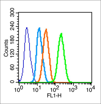
Blank control (blue line): Hela (blue). Primary Antibody (green line): Rabbit Anti-Connexin 43 antibody (orb10485), Dilution: 1 µg/10 6 cells, Isotype Control Antibody (orange line): Rabbit IgG. Secondary Antibody (white blue line): F (ab)2 fragment goat anti-rabbit IgG-FITC. Dilution: 1 µg/Test. Protocol, The cells were fixed with 2% paraformaldehyde (10 min), then permeabilized with 90% ice-cold methanol for 30 min on ice. Cells stained with Primary Antibody for 30 min at room temperature. The cells were then incubated in 1 X PBS/2% BSA/10% goat serum to block non-specific protein-protein interactions followed by the antibody for 15 min at room temperature. The secondary antibody used for 40 min at room temperature. Acquisition of 20000 events was performed. |
|
|
Paraformaldehyde-fixed, paraffin embedded (Human stomach cancer), Antigen retrieval by boiling in sodium citrate buffer (pH6.0) for 15 min, Block endogenous peroxidase by 3% hydrogen peroxide for 20 minutes, Blocking buffer (normal goat serum) at 37C for 30 min, Antibody incubation with (Connexin 43) Polyclonal Antibody, Unconjugated (orb10485) at 1:200 overnight at 4C, followed by operating according to SP Kit (Rabbit) instructions and DAB staining. |
|
|
Paraformaldehyde-fixed, paraffin embedded (Mouse brain), Antigen retrieval by boiling in sodium citrate buffer (pH6.0) for 15 min, Block endogenous peroxidase by 3% hydrogen peroxide for 20 minutes, Blocking buffer (normal goat serum) at 37C for 30 min, Antibody incubation with (Connexin 43) Polyclonal Antibody, Unconjugated (orb10485) at 1:400 overnight at 4C, followed by operating according to SP Kit (Rabbit) instructionsand DAB staining. |
|
|
Sample: Lane 1: Heart (Mouse) Lysate at 40 ug, Lane 2: Heart (Rat) Lysate at 40 ug, Lane 3: Cerebrum (Mouse) Lysate at 40 ug, Lane 4: Cerebrum (Rat) Lysate at 40 ug, Primary: Anti-Connexin 43 (orb10485) at 1/1000 dilution, Secondary: IRDye800CW Goat Anti-Rabbit IgG at 1/20000 dilution, Predicted band size: 42 kD, Observed band size: 45 kD. |
|
|
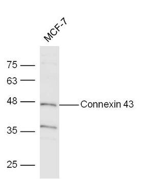
Sample: Mcf-7 Cell Lysate at 40 ug, Primary: Anti-Connexin (orb10485) at 1/300 dilution, Secondary: IRDye800CW Goat Anti-Rabbit IgG at 1/10000 dilution, Predicted band size: 42 kD, Observed band size: 43 kD. |
|
|
Tissue/Cell: MCF7 cell, 4% Paraformaldehyde-fixed, Triton X-100 at room temperature for 20 min, Blocking buffer (normal goat serum) at 37C for 20 min, Antibody incubation with (Connexin 43) polyclonal Antibody, Unconjugated (orb10485) 1:100, 90 minutes at 37C, followed by a FITC conjugated Goat Anti-Rabbit IgG antibody at 37C for 90 minutes, DAPI (blue) was used to stain the cell nuclei. |
|
|
Tissue/Cell: U-251 cell, 4% Paraformaldehyde-fixed, Triton X-100 at room temperature for 20 min, Blocking buffer (normal goat serum) at 37C for 20 min, Antibody incubation with (Connexin 43) polyclonal Antibody, Unconjugated (orb10485) 1:100, 90 minutes at 37C, followed by a FITC conjugated Goat Anti-Rabbit IgG antibody at 37C for 90 minutes, DAPI (blue) was used to stain the cell nuclei. |
Product Guarantee and Expert Support
