![]()
|
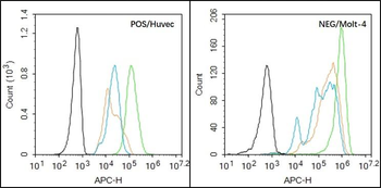
Black line: Positive blank control (HUVEC), Negative blank control (Molt-4), Green line: Primary Antibody (Rabbit Anti-EGFR antibody (orb10580)), Orange line: Isotype Control Antibody (Rabbit IgG). Blue line: Secondary Antibody (Goat anti-rabbit IgG-AF647), HUVEC (Positive) and Molt-4 (Negative control) cells (black) were fixed with 4% PFA for 10 min at room temperature, permeabilized with 90% ice-cold methanol for 20 min at -20C, and incubated in 5% BSA blocking buffer for 30 min at room temperature. Cells were then stained with EGFR Antibody (orb10580) at 1:50 dilution in blocking buffer and incubated for 30 min at room temperature, washed twice with 2% BSA in PBS, followed by secondary antibody (blue) incubation for 40 min at room temperature. Acquisitions of 20000 events were performed. Cells stained with primary antibody (green), and isotype control (orange). |
![]()
|
Blank control (blue line): A431 (blue). Primary Antibody (green line): Rabbit Anti-EGFRantibody (orb10580), Dilution: 3 µg/10 6 cells, Isotype Control Antibody (orange line): Rabbit IgG. Secondary Antibody (white blue line): Goat anti-rabbit IgG-FITC, Dilution: 1 µg/Test. Protocol, The cells were fixed with 2% paraformaldehyde (10 min), then permeabilized with 90% ice-cold methanol for 30 min on ice. Cells stained with Primary Antibody for 30 min at room temperature. The cells were then incubated in 1 X PBS/2% BSA/10% goat serum to block non-specific protein-protein interactions followed by the antibody for 15 min at room temperature. The secondary antibody used for 40 min at room temperature. Acquisition of 20000 events was performed. |
![]()
|
Blank control: HUVEC cells (blue). Primary Antibody: Rabbit Anti-EGFR antibody (orb10580), Dilution: 1 µg in 100 µl 1X PBS containing 0.5% BSA, Isotype Control Antibody: Rabbit IgG (orange), used under the same conditions, Secondary Antibody: Goat anti-rabbit IgG-PE (white blue), Dilution: 1:200 in 1 X PBS containing 0.5% BSA. Protocol, The cells were fixed with 2% paraformaldehyde (10 min), then permeabilized with 90% ice-cold methanol for 30 min on ice. Primary antibody (orb10580, 1 µg/1x10 6 cells) were incubated for 30 min on the ice, followed by 1 X PBS containing 0.5% BSA + 10% goat serum (15 min) to block non-specific protein-protein interactions. Then the Goat Anti-rabbit IgG/PE antibody was added into the blocking buffer mentioned above to react with the primary antibody at 1/200 dilution for 30 min on ice. Acquisition of 20000 events was performed. |
![]()
|
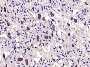
Paraformaldehyde-fixed, paraffin embedded (human gastric carcinoma), Antigen retrieval by boiling in sodium citrate buffer (pH6.0) for 15 min, Block endogenous peroxidase by 3% hydrogen peroxide for 20 minutes, Blocking buffer (normal goat serum) at 37C for 30 min, Antibody incubation with (EGFR) Polyclonal Antibody, Unconjugated (orb10580) at 1:200 overnight at 4C, followed by operating according to SP Kit (Rabbit) instructionsand DAB staining. |
![]()
|
Paraformaldehyde-fixed, paraffin embedded (rat placenta), Antigen retrieval by boiling in sodium citrate buffer (pH6.0) for 15 min, Block endogenous peroxidase by 3% hydrogen peroxide for 20 minutes, Blocking buffer (normal goat serum) at 37C for 30 min, Antibody incubation with (EGFR) Polyclonal Antibody, Unconjugated (orb10580) at 1:200 overnight at 4C, followed by operating according to SP Kit (Rabbit) instructionsand DAB staining. |
![]()
|
Sample: Lane 1: Hela (Human) Cell Lysate at 30 ug, Lane 2: A549 (Human) Cell Lysate at 30 ug, Lane 3: U251 (Human) Cell Lysate at 30 ug, Lane 4: U87MG (Human) Cell Lysate at 30 ug, Primary: Anti-EGFR at 1/1000 dilution, Secondary: IRDye800CW Goat Anti-Rabbit IgG at 1/20000 dilution, Predicted band size: 170 kD, Observed band size: 170 kD. |
![]()
|
Tissue/Cell: human rectal carcinoma, 4% Paraformaldehyde-fixed and paraffin-embedded, Antigen retrieval: citrate buffer (0.01M, pH6.0), Boiling bathing for 15 min, Blocking buffer (normal goat serum) at 37C for 20 min, Incubation: Anti-EGFR Polyclonal Antibody, Unconju |