
ERK1 + ERK2 Rabbit Polyclonal Antibody, Unconjugated
Catalog Number:
BYT-ORB10604
- Images (6)


| Article Name: | ERK1 + ERK2 Rabbit Polyclonal Antibody, Unconjugated |
| Biozol Catalog Number: | BYT-ORB10604 |
| Supplier Catalog Number: | orb10604 |
| Alternative Catalog Number: | BYT-ORB10604-50,BYT-ORB10604-100,BYT-ORB10604-200 |
| Manufacturer: | Biorbyt |
| Host: | Rabbit |
| Category: | Antikörper |
| Application: | FC, IF, IHC-Fr, IHC-P, WB |
| Species Reactivity: | Human, Mouse, Rat |
| Immunogen: | KLH conjugated synthetic peptide derived from human ERK2 (301-358/358aa) |
| Conjugation: | Unconjugated |
| Alternative Names: | MK03_HUMAN, MAPK3, MAP kinase 3, MAPK 3, ERT2, Extracellular signal-regulated kinase 1 (ERK-1), Insulin-stimulated MAP2 kinase, MAP kinase isoform p44 (p44-MAPK), Microtubule-associated protein 2 kinase, p44-ERK1, 2.7.11.24, ERK1, PRKM3, MK01_HUMAN, MAPK1, MAP kinase 1, MAPK 1, ERT1, Extracellular signal-regulated kinase 2 (ERK-2), MAP kinase isoform p42 (p42-MAPK), Mitogen-activated protein kinase 2 (MAP kinase 2 | MAPK 2), ERK2, PRKM1, PRKM2, ERK, ERK-2, MAPK2, NS13, P42MAPK, p38, p40, p41, p41mapk, p42-MAPK, ERK-1, HS44KDAP, HUMKER1A, P44ERK1, P44MAPK, p44-MAPK, |
| Rabbit Anti-ERK1 + ERK2 antibody |
| Application Dilute: | WB=1:500-2000, IHC-P=1:100-500, IHC-F=1:100-500, IF=1:100-500, Flow-Cyt=1µg/Test |
|
|
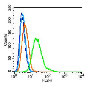
Blank control: Hep G2 cells (blue). Primary Antibody: Rabbit Anti-ERK1 + ERK2 antibody (orb10604), Dilution: 1 µg in 100 µl 1X PBS containing 0.5% BSA, Isotype Control Antibody: Rabbit IgG (orange), used under the same conditions, Secondary Antibody: Goat anti-rabbit IgG-PE (white blue), Dilution: 1:200 in 1 X PBS containing 0.5% BSA. Protocol, The cells were fixed with 2% paraformaldehyde (10 min), then permeabilized with 90% ice-cold methanol for 30 min on ice. Primary antibody (orb10604, 1 µg/1x10 6 cells) were incubated for 30 min on the ice, followed by 1 X PBS containing 0.5% BSA + 1 0% goat serum (15 min) to block non-specific protein-protein interactions. Then the Goat Anti-rabbit IgG/PE antibody was added into the blocking buffer mentioned above to react with the primary antibody at 1/200 dilution for 30 min on ice. Acquisition of 20000 events was performed. |
|
|
Paraformaldehyde-fixed, paraffin embedded (Mouse brain), Antigen retrieval by boiling in sodium citrate buffer (pH6.0) for 15 min, Block endogenous peroxidase by 3% hydrogen peroxide for 20 minutes, Blocking buffer (normal goat serum) at 37C for 30 min, Antibody incubation with (ERK1 + ERK2) Polyclonal Antibody, Unconjugated (orb10604) at 1:400 overnight at 4C, followed by operating according to SP Kit (Rabbit) instructionsand DAB staining. |
|
|
Sample: Brain (Rat) Lysate at 30 ug, Heart (Rat) lysate at 30 ug, Primary: Anti-ERK2/MAPK1 (orb10604) at 1/200 dilution, Secondary: HRP conjugated Goat-Anti-rabbit IgG (orb572747) at 1/3000 dilution, Predicted band size: 42 kD, Observed band size: 42 kD. |
|
|
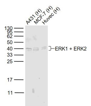
Sample: Lane 1: A431 (Human) Cell Lysate at 30 ug, Lane 2: MCF-7 (Human) Cell Lysate at 30 ug, Lane 3: Huvec (Human) Cell Lysate at 30 ug, Primary: Anti-ERK1 + ERK2 (orb10604) at 1/1000 dilution, Secondary: IRDye800CW Goat Anti-Rabbit IgG at 1/20000 dilution, Predicted band size: 44/42 kD, Observed band size: 42/40 kD. |
|
|
Sample: Lane 1: Cerebrum (Mouse) Lysate at 40 ug, Lane 2: Cerebrum (Rat) Lysate at 40 ug, Lane 3: Lymph node (Mouse) Lysate at 40 ug, Lane 4: Lymph node (Rat) Lysate at 40 ug, Primary: Anti-ERK1 + ERK2 (orb10604) at 1/1000 dilution, Secondary: IRDye800CW Goat Anti-Rabbit IgG at 1/20000 dilution, Predicted band size: 44/42 kD, Observed band size: 40 kD. |
|
|
Tissue/Cell: human lung carcinoma, 4% Paraformaldehyde-fixed and paraffin-embedded, Antigen retrieval: citrate buffer (0.01M, pH6.0), Boiling bathing for 15 min, Block endogenous peroxidase by 3% Hydrogen peroxide for 30 min, Blocking buffer (normal goat serum) at 37C for 20 min, Incubation: Anti-ERK2/MAPK1 Polyclonal Antibody, Unconjugated (orb10604) 1:200, overnight at 4C, followed by conjugation to the secondary antibody and DAB staining. |
Product Guarantee and Expert Support
