
Phospho-GSK-3 Beta (Ser9) Rabbit Polyclonal Antibody, Unconjugated
Catalog Number:
BYT-ORB10755
- Images (7)


| Article Name: | Phospho-GSK-3 Beta (Ser9) Rabbit Polyclonal Antibody, Unconjugated |
| Biozol Catalog Number: | BYT-ORB10755 |
| Supplier Catalog Number: | orb10755 |
| Alternative Catalog Number: | BYT-ORB10755-50,BYT-ORB10755-100,BYT-ORB10755-200 |
| Manufacturer: | Biorbyt |
| Host: | Rabbit |
| Category: | Antikörper |
| Application: | FC, ICC, IF, IHC-Fr, IHC-P, WB |
| Species Reactivity: | Human, Mouse, Rat |
| Immunogen: | KLH conjugated Synthesised phosphopeptide derived from human GSK-3 Beta around the phosphorylation site of Ser9 TT(p-S)FA |
| Conjugation: | Unconjugated |
| Alternative Names: | GSK3B | GSK3 beta (p-S9), p-GSK3 beta, phospho-GSK3 beta, 7330414F15Rik, 8430431H08Rik, GSK-3, GSK-3beta, GSK3, GSK3B_HUMAN, GSK3B, GSK-3 beta, Serine/threonine-protein kinase GSK3B, 2.7.11.26, GSK3B_MOUSE, |
| Phospho-GSK-3 Beta (Ser9) Rabbit Polyclonal Antibody |
| Application Dilute: | WB=1:500-2000, IHC-P=1:100-500, IHC-F=1:100-500, ICC/IF=1:100-500, IF=1:100-500, Flow-Cyt=1ug/Test |
| Application Notes: | Modification: Phosphorylated |
|
|
Blank control: A431. Primary Antibody (green line): Rabbit Anti-phospho-GSK-3 Beta (Ser9) antibody (orb10755), Dilution: 1 µg/10 6 cells, Isotype Control Antibody (orange line): Rabbit IgG. Secondary Antibody: Goat anti-rabbit IgG-AF488, Dilution: 1 µg/Test. Protocol, The cells were fixed with 4% PFA (10 min at room temperature) and then permeabilized with 90% ice-cold methanol for 20 min at -20C. The cells were then incubated in 5% BSA to block non-specific protein-protein interactions for 30 min at room temperature. Cells stained with Primary Antibody for 30 min at room temperature. The secondary antibody used for 40 min at room temperature. Acquisition of 20000 events was performed. |
|
|
Paraformaldehyde-fixed, paraffin embedded (Mouse placenta), Antigen retrieval by boiling in sodium citrate buffer (pH6.0) for 15 min, Block endogenous peroxidase by 3% hydrogen peroxide for 20 minutes, Blocking buffer (normal goat serum) at 37C for 30 min, Antibody incubation with (Beta (Ser9)) Polyclonal Antibody, Unconjugated at 1:500 overnight at 4C, followed by a conjugated secondary for 20 minutes and DAB staining. |
|
|
Sample: Lane 1: Mouse Cerebrum tissue lysates, Lane 2: Rat Cerebrum tissue lysates, Primary: Anti-phospho-GSK-3 Beta (Ser9) (orb10755) at 1/1000 dilution, Secondary: IRDye800CW Goat Anti-Rabbit IgG at 1/20000 dilution, Predicted band size: 47 kDa, Observed band size: 47 kDa. |
|
|
Sample: Testis (Rat) Lysate at 30 ug, Primary: Anti-phospho-GSK-3 Beta (Ser9) (orb10755) at 1:200 dilution, Secondary: HRP conjugated Goat Anti-Rabbit IgG (orb572747) at 1:5000 dilution, Predicted band size: 47kD, Observed band size: 49kD. |
|
|
The blue histogram is unstained cells (A549 cells). The Orange histogram is cells stained with Rabbit IgG/FITC (orb525556). The green histogram is cells stained with Rabbit Anti-phospho-GSK-3 Beta (Ser9)/FITC Conjugated antibody. Isotype control: Cell lines treated with Rabbit IgG/FITC (orb525556) instead of the primary antibody to confirm that primary antibody binding is specific. Concentration: 5 µl in 100 µl 1 X PBS containing 0.5% BSA. |
|
|
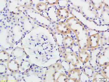
Tissue/Cell: rat kidney tissue, 4% Paraformaldehyde-fixed and paraffin-embedded, Antigen retrieval: citrate buffer (0.01M, pH6.0), Boiling bathing for 15 min, Block endogenous peroxidase by 3% Hydrogen peroxide for 30 min, Blocking buffer (normal goat serum) at 37C for 20 min, Incubation: Anti-phospho-GSK-3 Beta (Ser9) Polyclonal Antibody, Unconjugated (orb10755) 1:200, overnight at 4C, followed by conjugation to the secondary antibody and DAB staining. |
|
|
Tissue/Cell: Hela cell, 4% Paraformaldehyde-fixed, Triton X-100 at room temperature for 20 min, Blocking buffer (normal goat serum) at 37C for 20 min, Antibody incubation with (phospho-GSK-3 Beta (Ser9)) polyclonal Antibody, Unconjugated (orb10755) 1:100, 90 minutes at 37C, followed by a FITC conjugated Goat Anti-Rabbit IgG antibody at 37C for 90 minutes, DAPI (blue) was used to stain the cell nuclei. |
Product Guarantee and Expert Support
