Supplied at 0.5 mg/ml in Tris saline, 0.02% sodium azide, pH 7.3 with 0.5% bovine serum albumin. Aliquot and store at -20C. Minimize freezing and thawing.
Form:
Liquid
Target:
CD274
Application Notes:
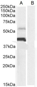
Application Notes: Peptide ELISA: antibody detection limit dilution 1:128000.Western Blot:Approx 40kDa band observed in Human Heart lysates. The observed molecular weight corresponds to glycosylation (calculated MW of 33.3kDa according to NP_054862.1). An additional band was also consistently observed at 55kDa, and was successfully blocked by incubation with the immunizing peptide. Preliminary testing also showed the 37+55kDa bands in lysates of cell lines, A549, Daudi, HeLa, HepG2 and Jurkat Recommended concentration: 0.03-0.1µg/mL. Primary incubation 1 hour at room temperature.Immunohistochemistry:In paraffin embedded Human Placenta shows membranous staining of cytotrophoblasts. Recommended concentration: 2-4µg/mL.Immunofluorescence: Strong expression of the protein seen in the cytoplasm of A431 and U2OS cells. Recommended concentration: 10µg/mL. Flow Cytometry: Flow cytometric analysis of Jurkat cells. Recommended concentration: 10µg/mL
orb1247125 (0.3 ug/ml) staining of Human Heart (A) lysate + Blocking peptide (B) (35 ug protein in RIPA buffer). Detected by chemiluminescence.
orb1247125 (2 ug/ml) staining of paraffin embedded Human Placenta. Microwaved antigen retrieval with citrate buffer pH 6, HRP-staining.
orb1247125 Negative Control showing staining of paraffin embedded Human Placenta, with no primary antibody.
orb1247125 Immunofluorescence analysis of paraformaldehyde fixed A431 cells, permeabilized with 0.15% Triton. Primary incubation 1hr (10 ug/ml) followed by Alexa Fluor 488 secondary antibody (2 ug/ml), showing cytoplasmic staining. The nuclear stain is DAPI.
orb1247125 Immunofluorescence analysis of paraformaldehyde fixed U2OS cells, permeabilized with 0.15% Triton. Primary incubation 1hr (10 ug/ml) followed by Alexa Fluor 488 secondary antibody (2 ug/ml), showing cytoplasmic staining. The nuclear stain is DAPI.
orb1247125 Flow cytometric analysis of paraformaldehyde fixed Jurkat cells (blue line), permeabilized with 0.5% Triton. Primary incubation 1hr (10 ug/ml) followed by Alexa Fluor 488 secondary antibody (1 ug/ml). IgG control: Unimmunized goat IgG (black line).
* VAT and and shipping costs not included. Errors and price changes excepted