PBS with 0.1 mg/ml rAlbumin and 0.05% sodium azide
Form:
Liquid
Target:
HSPD1
Application Notes:
Application Notes: Flow Cytometry: 0.5-1 ug/million cellsIF: 0.5-1 µg/mLWB: 0.25-0.5 µg/mLIHC (FFPE): 0.5-1 µg/mL for 30 minutes at RT (1)Prediluted format : incubate for 30 min at RT (2)The concentration stated for each application is a general starting point. Variations in protocols, secondaries and substrates may require the antibody to be titered up or down for optimal performance.1. Staining of formalin-fixed tissues is enhanced by boiling tissue sections in 10mM Citrate Buffer, pH 6.0, for 10-20 min followed by cooling at RT for 20 minutes.2. The prediluted format is supplied in a dropper bottle and is optimized for use in IHC. After epitope retrieval step (if required), drip mAb solution onto the tissue section and incubate at RT for 30 min
Formalin/paraffin human breast carcinoma stained with HSP60 antibody.
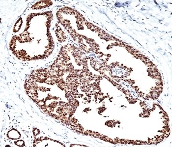
IHC testing of FFPE pancreas tissue with HSP60 antibody.
Western blot testing of HeLa cell lysate with HSP60 antibody (clone LK1).
SDS-PAGE Analysis of Purified, BSA-Free HSP60 Antibody (clone LK1). Confirmation of Integrity and Purity of the Antibody.
Immunofluorescent staining of PFA-fixed human MCF7 cells with HSP60 antibody (green, clone LK1) and Reddot nuclear stain (red).
Flow cytometry testing of PFA-fixed human HeLa cells with HSP60 antibody (clone LK1), Red = isotype control, Blue = HSP60 antibody.
* VAT and and shipping costs not included. Errors and price changes excepted