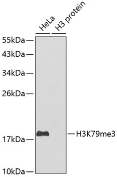
Western blot analysis of extracts of various cell lines, using TriMethyl-Histone H3-K79 antibody (orb1258179). Secondary antibody: HRP Goat Anti-Rabbit IgG (H+L) at 1:10000 dilution. Lysates/proteins: 25 ug per lane. Blocking buffer: 3% nonfat dry milk in TBST.
Dot-blot analysis of all sorts of methylation peptides using TriMethyl-Histone H3-K79 antibody (orb1258179) at 1:1000 dilution.
Immunohistochemistry of paraffin-embedded human mammary cancer using TriMethyl-Histone H3-K79 antibody (orb1258179) at dilution of 1:200 (40x lens).
Immunohistochemistry of paraffin-embedded rat testis using TriMethyl-Histone H3-K79 antibody (orb1258179) at dilution of 1:200 (40x lens).
Immunohistochemistry of paraffin-embedded mouse testis using TriMethyl-Histone H3-K79 antibody (orb1258179) at dilution of 1:200 (40x lens).
Immunofluorescence analysis of 293T cells using TriMethyl-Histone H3-K79 antibody (orb1258179). Blue: DAPI for nuclear staining.
Chromatin immunoprecipitation analysis of extracts of 293 cell line, using H3K79me3 antibody (orb1258179) and rabbit IgG. The amount of immunoprecipitated DNA was checked by quantitative PCR. Histogram was constructed by the ratios of the immunoprecipitated DNA to the input.
* VAT and and shipping costs not included. Errors and price changes excepted