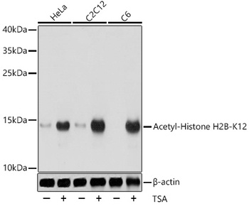
Western blot analysis of extracts of various cell lines, using Acetyl-Histone H2B-K12 antibody (orb1259423) at 1:1000 dilution. HeLa cells were treated by TSA (1 uM) at 37 °C for 18 hours. C2C12 cells were treated by TSA (1 uM) at 37 °C for 18 hours. C6 cells were treated by TSA (1 uM) at 37 °C for 18 hours. Secondary antibody: HRP Goat Anti-Rabbit IgG (H+L) at 1:10000 dilution. Lysates/proteins: 25 ug per lane. Blocking buffer: 3% BSA. Detection: ECL Basic Kit. Exposure time: 1s.
Immunohistochemistry of paraffin-embedded human colon using Acetyl-Histone H2B-K12 antibody (orb1259423) at dilution of 1:200 (40x lens).
Immunohistochemistry of paraffin-embedded rat ovary using Acetyl-Histone H2B-K12 antibody (orb1259423) at dilution of 1:200 (40x lens).
Immunohistochemistry of paraffin-embedded mouse testis using Acetyl-Histone H2B-K12 antibody (orb1259423) at dilution of 1:200 (40x lens).
Immunofluorescence analysis of C6 cells using Acetyl-Histone H2B-K12 antibody (orb1259423) at dilution of 1:100. C6 cells were treated by TSA (1 uM) at 37 °C for 18 hours. Blue: DAPI for nuclear staining.
Immunofluorescence analysis of HeLa cells using Acetyl-Histone H2B-K12 antibody (orb1259423) at dilution of 1:100. HeLa cells were treated by TSA (1 uM) at 37 °C for 18 hours. Blue: DAPI for nuclear staining.
Immunofluorescence analysis of NIH/3T3 cells using Acetyl-Histone H2B-K12 antibody (orb1259423) at dilution of 1:100. NIH/3T3 cells were treated by TSA (1 uM) at 37 °C for 18 hours. Blue: DAPI for nuclear staining.
* VAT and and shipping costs not included. Errors and price changes excepted