
CTCF Antibody, Unconjugated, Rabbit, Polyclonal
Catalog Number:
BYT-ORB1260507
- Images (8)


| Article Name: | CTCF Antibody, Unconjugated, Rabbit, Polyclonal |
| Biozol Catalog Number: | BYT-ORB1260507 |
| Supplier Catalog Number: | orb1260507 |
| Alternative Catalog Number: | BYT-ORB1260507-100 |
| Manufacturer: | Biorbyt |
| Host: | Rabbit |
| Category: | Antikörper |
| Application: | ChIP, IF, IHC, IP, WB |
| Species Reactivity: | Human, Mouse, Rat |
| Immunogen: | Recombinant fusion protein containing a sequence corresponding to amino acids 1-260 of human CTCF (NP_006556.1). |
| Conjugation: | Unconjugated |
| Alternative Names: | CTCF, MRD21 |
| CTCF Antibody |
| Clonality: | Polyclonal |
| Concentration: | batch dependent |
| Molecular Weight: | Observed: 140kDa |
| UniProt: | P49711 |
| Buffer: | PBS with 0.02% sodium azide, 50% glycerol, pH 7.3. |
| Form: | Liquid |
| Target: | CTCF |
| Application Notes: | Application Notes: WB: 1:1000 - 1:2000IHC: 1:50 - 1:200IF: 1:50 - 1:200IP: 1:50 - 1:100ChIP: 1:50 - 1:200 |
|
|
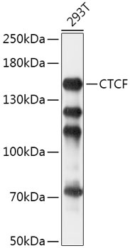
Western blot analysis of extracts of 293T cells, using CTCF antibody (orb1260507) at 1:1000 dilution. Secondary antibody: HRP Goat Anti-Rabbit IgG (H+L) at 1:10000 dilution. Lysates/proteins: 25 ug per lane. Blocking buffer: 3% nonfat dry milk in TBST. Detection: ECL Basic Kit. Exposure time: 5s. |
|
|
Immunohistochemistry of paraffin-embedded rat liver using CTCF antibody (orb1260507) at dilution of 1:200 (40x lens). |
|
|
Immunohistochemistry of paraffin-embedded rat brain using CTCF antibody (orb1260507) at dilution of 1:200 (40x lens). |
|
|
Immunohistochemistry of paraffin-embedded rat spleen using CTCF antibody (orb1260507) at dilution of 1:200 (40x lens). |
|
|
Immunohistochemistry of paraffin-embedded mouse brain using CTCF antibody (orb1260507) at dilution of 1:200 (40x lens). |
|
|
Immunofluorescence analysis of U2OS cells using CTCF antibody (orb1260507) at dilution of 1:100. Blue: DAPI for nuclear staining. |
|
|
Immunoprecipitation analysis of 200 ug extracts of HeLa cells, using 3 ug CTCF antibody (orb1260507). Western blot was performed from the immunoprecipitate using CTCF antibody (orb1260507) at a dilution of 1:1000. |
|
|
Chromatin immunoprecipitation analysis of extracts of HCT116 cells, using CTCF antibody (orb1260507) and rabbit IgG. The amount of immunoprecipitated DNA was checked by quantitative PCR. Histogram was constructed by the ratios of the immunoprecipitated DNA to the input. |
Product Guarantee and Expert Support
