Synthetic peptide / encompassing a sequence within the center region
Conjugation:
Unconjugated
Rabbit polyclonal antibody to Cytokeratin 14
Clonality:
Polyclonal
Buffer:
100mM Tris Glycine, 3% rAlbumin, 20% Glycerol (pH7). 0.025% ProClin 300 was added as a preservative
Target:
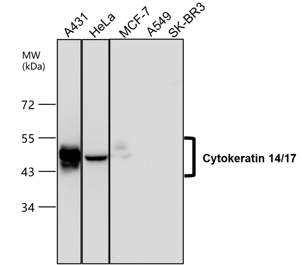
Cytokeratin 14/17
Application Dilute:
Western Blot 1:1000-1:2000, Immunofluorescence 1:100-1:400, Immunohistochemistry (Paraffin) 1:100-1:300
All lanes: Anti-Cytokeratin 14/17 antibody - at 1/2000 dilution, Lysates/proteins at 45 µg per lane. This blot was produced using a 10% SDS-PAGE. The gel was run at 140V for 50 minutes before being transferred onto a Nitrocellulose membrane at 18V for 60 minutes. The membrane was then blocked an hour before being incubated with orb1294304 overnight at 4C.
Immunofluorescence: cells were fixed with 4% paraformaldehyde for 10 min at RT, permeabilized with 0.1% NP-40 for 10 min at RT then blocked with 5% BSA for 30 min at room temperature. Cells were stained with orb1294304 anti-Cytokeratin 14/17 antibody (red) at 1:150 and 4C. DAPI (blue) was used as the nuclear counter stain.
Immunofluorescence: cells were fixed with 4% paraformaldehyde for 10 min at RT, permeabilized with 0.1% NP-40 for 10 min at RT then blocked with 5% BSA for 30 min at room temperature. Cells were stained with orb1294304 anti-Cytokeratin 14/17 antibody (red) at 1:150 and 4C. DAPI (blue) was used as the nuclear counter stain.
Immunohistochemical analysis of paraffin embedded Human cancer tissue labeling Cytokeratin 14/17 with orb1294304 at 1/100.
Immunohistochemical analysis of paraffin embedded Human cancer tissue labeling Cytokeratin 14/17 with orb1294304 at 1/100.
Immunohistochemical analysis of paraffin embedded Human cancer tissue labeling Cytokeratin 14/17 with orb1294304 at 1/100.
Immunohistochemical analysis of paraffin embedded Human cancer tissue labeling Cytokeratin 14/17 with orb1294304 at 1/100.
* VAT and and shipping costs not included. Errors and price changes excepted