
P53 (wt-p53) Rabbit Polyclonal Antibody, Unconjugated
Catalog Number:
BYT-ORB13631
- Images (6)


| Article Name: | P53 (wt-p53) Rabbit Polyclonal Antibody, Unconjugated |
| Biozol Catalog Number: | BYT-ORB13631 |
| Supplier Catalog Number: | orb13631 |
| Alternative Catalog Number: | BYT-ORB13631-50,BYT-ORB13631-100,BYT-ORB13631-200 |
| Manufacturer: | Biorbyt |
| Host: | Rabbit |
| Category: | Antikörper |
| Application: | ICC, WB |
| Species Reactivity: | Human |
| Immunogen: | KLH conjugated synthetic peptide derived from human P53 (301-393/393aa) |
| Conjugation: | Unconjugated |
| Alternative Names: | BCC7, BMFS5, LFS1, P53, TRP53, Tp53, bbl, bfy, bhy, p44, P53_BOVIN, Tumor suppressor p53, P53_HUMAN, Antigen NY-CO-13, Phosphoprotein p53, P53_MOUSE, P53_RAT, |
| P53 (wt-p53) Rabbit Polyclonal Antibody |
| Clonality: | Polyclonal |
| Concentration: | 1mg/ml |
| Molecular Weight: | 53 kDa |
| UniProt: | P04637 |
| Buffer: | 0.01M TBS (pH7.4) with 1% rAlbumin, 0.02% Proclin300 and 50% Glycerol. |
| Form: | Liquid |
| Target: | TP53 |
| Application Dilute: | WB=1:500-2000, ICC/IF=1:100-500 |
|
|
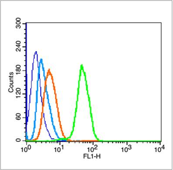
Blank control (blue line): Hela (blue). Primary Antibody (green line): Rabbit Anti-P53 (wt-p53) antibody (orb13631), Dilution: 1 µg/10 6 cells, Isotype Control Antibody (orange line): Rabbit IgG. Secondary Antibody (white blue line): Goat anti-rabbit IgG-FITC, Dilution: 1 µg/Test. Protocol, The cells were fixed with 70% ethanol (Overnight at 4C) and then permeabilized with 90% ice-cold methanol for 30 min on ice. Cells stained with Primary Antibody for 30 min at room temperature. The cells were then incubated in 1X PBS/2% BSA/10% goat serum to block non-specific protein-protein interactions followed by the antibody for 15 min at room temperature. The secondary antibody used for 40 min at room temperature. Acquisition of 20000 events was performed. |
|
|
Sample: Liver (Mouse) Lysate at 40 ug, Liver (Rat) Lysate at 40 ug, Primary: Anti-P53 (wt-p53) (orb13631) at 1/300 dilution, Secondary: IRDye800CW Goat Anti-Rabbit IgG at 1/20000 dilution, Predicted band size: 53 kD, Observed band size: 53 kD. |
|
|
Sample: MCF-7 (Human) Cell Lysate at 40 ug, DU145 (Human) Cell Lysate at 40 ug, Primary: Anti-P53 (wt-p53) (orb13631) at 1/300 dilution, Secondary: IRDye800CW Goat Anti-Rabbit IgG at 1/20000 dilution, Predicted band size: 53 kD, Observed band size: 53 kD. |
|
|
Tissue/Cell: A549 cell, 4% Paraformaldehyde-fixed, Triton X-100 at room temperature for 20 min, Blocking buffer (normal goat serum) at 37C for 20 min, Antibody incubation with (P53 (wt-p53)) polyclonal Antibody, Unconjugated (orb13631) 1:100, 90 minutes at 37C, followed by a FITC conjugated Goat Anti-Rabbit IgG antibody at 37C for 90 minutes, DAPI (blue) was used to stain the cell nuclei. |
|
|
Tissue/Cell: rat brain tissue, 4% Paraformaldehyde-fixed and paraffin-embedded, Antigen retrieval: citrate buffer (0.01M, pH 6.0), Boiling bathing for 15 min, Block endogenous peroxidase by 3% Hydrogen peroxide for 30 min, Blocking buffer (normal goat serum) at 37C for 20 min, Incubation: Anti-P53 (wt-p53) Polyclonal Antibody, Unconjugated (orb13631) 1:200, overnight at 4C, followed by conjugation to the secondary antibody and DAB staining. |
Product Guarantee and Expert Support
