0.01M TBS (pH7.4) with 1% rAlbumin, 0.02% Proclin300 and 50% Glycerol.
Form:
Liquid
Target:
PDCD1
Application Dilute:
Flow-Cyt=1µg /test
Immunohistochemical analysis of paraffin-embedded human cervical cancer tissue using PD1 antibody
IHC-P of human cervical cancer tissue (PD1 antibody at 1:300)
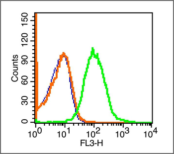
Blank control (blue line): Mouse spleen cells (blue). Primary Antibody (green line): Rabbit Anti-PD-1/PE-CY7 Conjugated antibody, Dilution: 1 µg/10 6 cells, Isotype Control Antibody (orange line): Rabbit IgG-PE-CY7. Protocol, The cells were fixed with 70% ice-cold methanol overnight at 4C. The cells were then incubated in 1X PBS/2% BSA/10% goat serum to block non-specific protein-protein interactions followed by the antibody for 15 min at room temperature. Cells stained with Primary Antibody for 30 min at room temperature. Acquisition of 20000 events was performed.
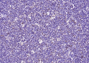
Paraformaldehyde-fixed, paraffin embedded (human tonsil), Antigen retrieval by boiling in sodium citrate buffer (pH6.0) for 15 min, Block endogenous peroxidase by 3% hydrogen peroxide for 20 minutes, Blocking buffer (normal goat serum) at 37C for 30 min, Antibody incubation with (PD-1) Polyclonal Antibody, Unconjugated (orb13641) at 1:200 overnight at 4C, followed by operating according to SP Kit (Rabbit) instructionsand DAB staining.
Paraformaldehyde-fixed, paraffin embedded (rat spleen), Antigen retrieval by boiling in sodium citrate buffer (pH6.0) for 15 min, Block endogenous peroxidase by 3% hydrogen peroxide for 20 minutes, Blocking buffer (normal goat serum) at 37C for 30 min, Antibody incubation with (PD-1) Polyclonal Antibody, Unconjugated (orb13641) at 1:200 overnight at 4C, followed by operating according to SP Kit (Rabbit) instructionsand DAB staining.
Sample: Hela (Human) Cell Lysate at 30 ug, MOLT-4 (Human) Cell Lysate at 30 ug, Jurkat (Human) Cell Lysate at 30 ug, Primary: Anti-PD-1 (orb13641) at 1/500 dilution, Secondary: IRDye800CW Goat Anti-Rabbit IgG at 1/20000 dilution, Predicted band size: 32 kD, Observed band size: 55 kD.
Sample: Raji (Human) Cell Lysate at 30 ug, Thymus (Mouse) Lysate at 40 ug, Lymph node (Mouse) Lysate at 40 ug, Spleen (Mouse) Lysate at 40 ug, Raw264.7 (Mouse) Cell Lysate at 40 ug, Thymus (Rat) Lysate at 40 ug, Primary: Anti-PD-1 (orb13641) at 1/1000 dilution, Secondary: IRDye800CW Goat Anti-Rabbit IgG at 1/20000 dilution, Predicted band size: 55 kD, Observed band size: 58 kD.
* VAT and and shipping costs not included. Errors and price changes excepted