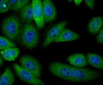
ICC staining Alcohol Dehydrogenase in HepG2 cells (green). The nuclear counter stain is DAPI (blue). Cells were fixed in paraformaldehyde, permeabilised with 0.25% Triton X100/PBS.
Immunohistochemical analysis of paraffin-embedded human liver tissue using anti-Alcohol Dehydrogenase antibody. Counter stained with hematoxylin.
Immunohistochemical analysis of paraffin-embedded mouse liver tissue using anti-Alcohol Dehydrogenase antibody. Counter stained with hematoxylin.
Immunohistochemical analysis of paraffin-embedded rat liver tissue using anti-Alcohol Dehydrogenase antibody. Counter stained with hematoxylin.
Sample: Lane 1: Rat Liver tissue lysates, Primary: Anti-ADH1A (orb1499353) at 1/500 dilution, Secondary: IRDye800CW Goat Anti-Rabbit IgG at 1/20000 dilution, Predicted band size: 41 kDa, Observed band size: 41 kDa.
* VAT and and shipping costs not included. Errors and price changes excepted