
BMAL1 Recombinant Rabbit Monoclonal Antibody, Clone: [8D1], Unconjugated
Catalog Number:
BYT-ORB1499364
- Images (6)


| Article Name: | BMAL1 Recombinant Rabbit Monoclonal Antibody, Clone: [8D1], Unconjugated |
| Biozol Catalog Number: | BYT-ORB1499364 |
| Supplier Catalog Number: | orb1499364 |
| Alternative Catalog Number: | BYT-ORB1499364-100,BYT-ORB1499364-25,BYT-ORB1499364-50 |
| Manufacturer: | Biorbyt |
| Host: | Rabbit |
| Category: | Antikörper |
| Application: | FC, WB |
| Species Reactivity: | Human, Mouse, Rat |
| Immunogen: | KLH conjugated synthetic peptide derived from human BMAL1 |
| Conjugation: | Unconjugated |
| Alternative Names: | ARNTL, ARNTL1, BMAL1c, JAP3, MOP3, PASD3, TIC, bHLHe5, Arnt3, BMAL1b, bmal1b, BMAL1_HUMAN, BMAL1, Aryl hydrocarbon receptor nuclear translocator-like protein 1, Basic-helix-loop-helix-PAS protein MOP3, Brain and muscle ARNT-like 1, Class E basic helix-loop-helix protein 5 (bHLHe5), Member of PAS protein 3, PAS domain-containing protein 3, bHLH-PAS protein JAP3, BMAL1_MOUSE, BMAL1_RAT, |
| BMAL1 Recombinant Rabbit Monoclonal Antibody |
| Clonality: | Recombinant |
| Concentration: | 1mg/ml |
| Clone Designation: | [8D1] |
| Molecular Weight: | 69 kDa |
| UniProt: | O00327 |
| Buffer: | 0.01M TBS (pH7.4) with 1% rAlbumin, 0.02% Proclin300 and 50% Glycerol. |
| Form: | Liquid |
| Target: | BMAL1 |
| Application Dilute: | WB=1:500-2000, Flow-Cyt=1:50-100 |
|
|
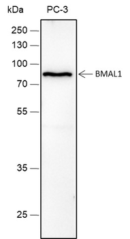
Blocking buffer: 5% NFDM/TBST, Primary ab Dilution: 1:2000, Primary ab incubation condition: 2 hours at room temperature, Secondary ab: Goat Anti-Rabbit IgG H&L (HRP), Lysate: PC-3, Protein loading quantity: 20 µg, Exposure time: 60 s, Predicted MW: 68 kDa, Observed MW: 75 kDa. |
|
|
Cell line: SH-SY5Y, Fixation: 4% Paraformaldehyde, Permeabilization: 90% Methanol, Primary Ab Dilution: 1:100, Secondary Ab: Goat Anti-Rabbit IgG, Unlabelled control: The cell without incubation with primary antibody and secondary antibody (Black line). Isotype control: Rabbit monoclonal IgG (Blue line). Comment: Line red is the positive signal for orb1499364. |
|
|
ICC staining of BMAL1 in Hela cells (green). Formalin fixed cells were permeabilized with 0.1% Triton X-100 in TBS for 10 minutes at room temperature and blocked with 1% Blocker BSA for 15 minutes at room temperature. Cells were probed with the primary antibody (orb1499364, 1/50) for 1 hour at room temperature, washed with PBS. Alexa Fluor488 Goat anti-Rabbit IgG was used as the secondary antibody at 1/1000 dilution. The nuclear counter stain is DAPI (blue). |
|
|
Tissue: Mouse hippocampus, Section type: Formalin fixed & Paraffin embedded section, Retrieval method: High temperature and high pressure, Retrieval buffer: Tris/EDTA buffer, pH9.0 Primary ab Dilution: 1:1000, Primary ab incubation condition: 1 hour at room temperature, Secondary ab: SP Kit (Rabbit), Counter stain: Hematoxylin (Blue), Comment: Color brown is the positive signal for orb1499364. |
|
|
Tissue: Mouse pancreas, Section type: Formalin fixed & Paraffin embedded section, Retrieval method: High temperature and high pressure, Retrieval buffer: Tris/EDTA buffer, pH9.0 Primary ab Dilution: 1:1000, Primary ab incubation condition: 1 hour at room temperature, Secondary ab: SP Kit (Rabbit), Counter stain: Hematoxylin (Blue), Comment: Color brown is the positive signal for orb1499364. |
|
|
Western blot analysis of BMAL1 on different lysates. Proteins were transferred to a PVDF membrane and blocked with 5% BSA in PBS for 1 hour at room temperature. The primary antibody (orb1499364, 1/500) was used in 5% BSA at room temperature for 2 hours. Goat Anti-Rabbit IgG - HRP Secondary Antibody (HA1001) at 1:5000 dilution was used for 1 hour at room temperature. Positive control: Lane 1: Hela cell lysate, Lane 2: NIH/3T3 cell lysate, Lane 3: Rat brain tissue lysate, Lane 4: Mouse spleen tissue lysate. |
Product Guarantee and Expert Support
