0.01M TBS (pH7.4) with 1% rAlbumin, 0.02% Proclin300 and 50% Glycerol.
Form:
Liquid
Target:
NOS1
Application Dilute:
WB=1:500-1000
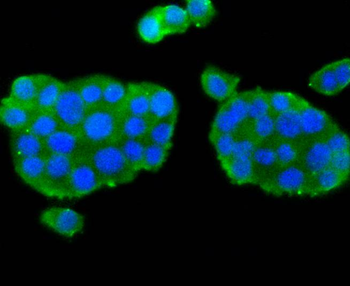
ICC staining of nNOS in PC-12 cells (green). Formalin fixed cells were permeabilized with 0.1% Triton X-100 in TBS for 10 minutes at room temperature and blocked with 1% Blocker BSA for 15 minutes at room temperature. Cells were probed with the primary antibody (orb1499394, 1/50) for 1 hour at room temperature, washed with PBS. Alexa Fluor488 Goat anti-Rabbit IgG was used as the secondary antibody at 1/1000 dilution. The nuclear counter stain is DAPI (blue).
ICC staining of nNOS in PC-3M cells (green). Formalin fixed cells were permeabilized with 0.1% Triton X-100 in TBS for 10 minutes at room temperature and blocked with 1% Blocker BSA for 15 minutes at room temperature. Cells were probed with the primary antibody (orb1499394, 1/50) for 1 hour at room temperature, washed with PBS. Alexa Fluor488 Goat anti-Rabbit IgG was used as the secondary antibody at 1/1000 dilution. The nuclear counter stain is DAPI (blue).
ICC staining of nNOS in SH-SY5Y cells (green). Formalin fixed cells were permeabilized with 0.1% Triton X-100 in TBS for 10 minutes at room temperature and blocked with 1% Blocker BSA for 15 minutes at room temperature. Cells were probed with the primary antibody (orb1499394, 1/50) for 1 hour at room temperature, washed with PBS. Alexa Fluor488 Goat anti-Rabbit IgG was used as the secondary antibody at 1/1000 dilution. The nuclear counter stain is DAPI (blue).
Immunohistochemical analysis of paraffin-embedded mouse brain tissue using anti-nNOS antibody. The section was pre-treated using heat mediated antigen retrieval with Tris-EDTA buffer (pH 8.0-8.4) for 20 minutes. The tissues were blocked in 5% BSA for 30 minutes at room temperature, washed with ddH2O and PBS, and then probed with the primary antibody (orb1499394, 1/50) for 30 minutes at room temperature. The detection was performed using an HRP conjugated compact polymer system. DAB was used as the chromogen. Tissues were counterstained with hematoxylin and mounted with DPX.
Immunohistochemical analysis of paraffin-embedded mouse heart tissue using anti-nNOS antibody. The section was pre-treated using heat mediated antigen retrieval with Tris-EDTA buffer (pH 8.0-8.4) for 20 minutes. The tissues were blocked in 5% BSA for 30 minutes at room temperature, washed with ddH2O and PBS, and then probed with the primary antibody (orb1499394, 1/50) for 30 minutes at room temperature. The detection was performed using an HRP conjugated compact polymer system. DAB was used as the chromogen. Tissues were counterstained with hematoxylin and mounted with DPX.
Immunohistochemical analysis of paraffin-embedded rat brain tissue using anti-nNOS antibody. The section was pre-treated using heat mediated antigen retrieval with Tris-EDTA buffer (pH 8.0-8.4) for 20 minutes. The tissues were blocked in 5% BSA for 30 minutes at room temperature, washed with ddH2O and PBS, and then probed with the primary antibody (orb1499394, 1/50) for 30 minutes at room temperature. The detection was performed using an HRP conjugated compact polymer system. DAB was used as the chromogen. Tissues were counterstained with hematoxylin and mounted with DPX.
Immunohistochemical analysis of paraffin-embedded rat testis tissue using anti-nNOS antibody. The section was pre-treated using heat mediated antigen retrieval with Tris-EDTA buffer (pH 8.0-8.4) for 20 minutes. The tissues were blocked in 5% BSA for 30 minutes at room temperature, washed with ddH2O and PBS, and then probed with the primary antibody (orb1499394, 1/50) for 30 minutes at room temperature. The detection was performed using an HRP conjugated compact polymer system. DAB was used as the chromogen. Tissues were counterstained with hematoxylin and mounted with DPX.
Sample: Lane 1: Mouse Cerebellum tissue lysates, Lane 2: Rat Cerebellum tissue lysates, Primary: Anti-nNOS (orb1499394) at 1/1000 dilution, Secondary: IRDye800CW Goat Anti-Rabbit IgG at 1/20000 dilution, Predicted band size: 130 kDa, Observed band size: 180 kDa.
* VAT and and shipping costs not included. Errors and price changes excepted